Japanese
English
- 有料閲覧
- Abstract 文献概要
- 1ページ目 Look Inside
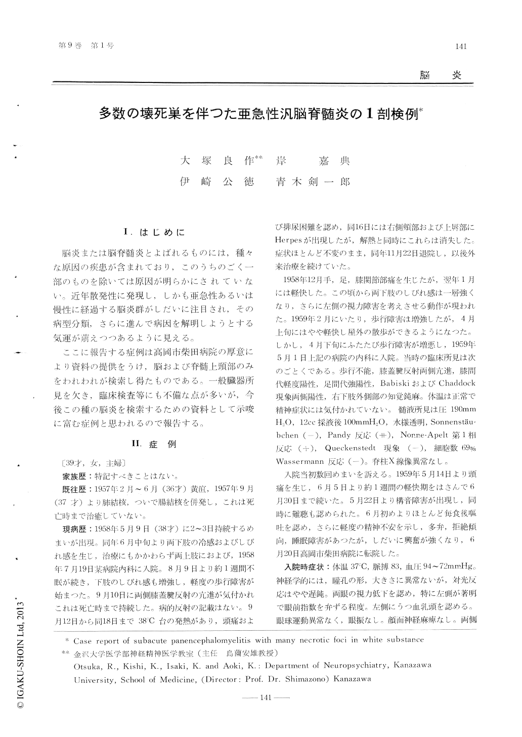
39才の主婦で,めまいや両下肢の知覚障害を初発症状として発病し,約2カ月後より軽度の歩行障害をしめし,4カ月後には一過性に発熱しHerpeslabiahsを生じている。約6ヵ月頃より視力障害さらに約1年後には精神症状や錐体路,錐体外路症状を認められるようになつて,しだいに病状は悪化し,全経過1年4カ月で死亡した。その間,脳脊髄液に軽度の細胞増多やグロブリン増加があり,急性播種性脳脊髄炎と考えられていた。
病理組織学的所見の特徴としては,終脳から脊髄にわたる,壊死傾向の強い白質の炎症性変化があげられる。すなわち大脳半球では後頭葉白質や脳梁膨大部など,半球後方部に粗大な壊死巣があり,血管周囲性の小円型細胞浸潤やミクログリアの増殖が強く,大脳脚や橋腕などにも同様の炎症像が認められる。小脳半球ではすでに瘢痕化した陳旧性の病巣が広範に存在している。しかし皮質神経細胞や脳幹諸核の神経細胞はほとんど侵されていない。また少数のグリア結節はみられたが,封入体細胞やNeuronophagieの像はみられない。
本症例は臨床病理学的に白質脳炎に近い性質をもつた亜急性壊死性汎脳脊髄炎であるが,従来の脳炎類型に例をみないものである。
The patient, a 39-year-old woman, had an onsetwith dizziness and hindrance of sensibility of boththe lower limbs in June 1958. About 2 monthsafter onset, she noticed slight dysbasia, and then 2months later, temporarily suffered from a feverand herpes labialis. About 6 months after onset, adisturbance of vision occurred. At the end of 10months of her illness, mental symptoms, pyramidal and extrapyramidal symptoms were observed.
From that time, the general conditions becamegradually aggravated, and she died after approximately 16 months of her illness.
The cerebrospinal fluid on admission showedslight pleocytosis and increased protein content. Our tentative clinical diagnosis was acute dissent-Mated encephalomyelitis.
After autopsy, the most marked pathological andanatomical findings were the presence of inflammatory foci in the white substance diffusing from thecerebral hemispheres to the spinal cord, and thesefoci had a tendency toward necrosis. The largenecrotic foci, the perivascular cuffs consisting oflymphocytes and an increased number of microgliawere found in the occipital lobe and the spleniumof the corpus callosum. Lesions of the cerebralwhite substance were usually most severe in theposterior part of the cerebrum and similar lesionswere detectable in the basis pedunculi and thebrachium pontis. In the cerebellar hemispheres,diffuse old cicatrical lesions were found. No changes were detected in nuclei of the cerebral corticesand of the basal ganglia. A few glial nodules werefound in the temporal white substance but nowherewere the inculsion body and the change of neuronophagy revealed.
We reported a case of Panen. cephalomyelitis necrotica subacuta. These clinical and pathologicalfeatures seem to resemble leucoencephalitis, but thepresent case is an encephalomyelitis not belongingto any of the classifications of encephalitides whichhave thus far been published.

Copyright © 1965, Igaku-Shoin Ltd. All rights reserved.


