Japanese
English
- 有料閲覧
- Abstract 文献概要
- 1ページ目 Look Inside
Ⅰ.まえがき
Nissl19)が1892年家兎の顔面神経を切断し,その神経核の病変を記載して以来,軸性変化に関する膨大な実験的研究がなされている14)。現在一般に知られている軸性変化の光学顕微鏡所見として,神経細胞体の腫脹,Niss1顆粒の崩壊,核の偏位,神経細胞周囲へのmicroglia集簇およびastrogliaの肥大が挙げられている10)。最近の運動神経核における軸性変化に関する微細組織研究では,神経細胞内のribosomeの増加と分散,小胞体消失,neurotubulesやneurofilamentの増加,Golgi apparatusの細胞周辺への分散および核小体空胞化が報告されている2)。
形態学的研究とともに軸性変化における神経細胞の機能および物質代謝も明らかにされつつある。軸性変化では,神経細胞内のRNAの増加とそれに伴う蛋白合成が亢進し4),同時に胞体内に新生された物質はaxonal flowに乗り正常時よりも多量に軸索末端に向かって輸送される17)といわれている。軸性変化の病理形態学とこれら物質代謝の変化の間に当然密接な関係が存在すると考えられる。
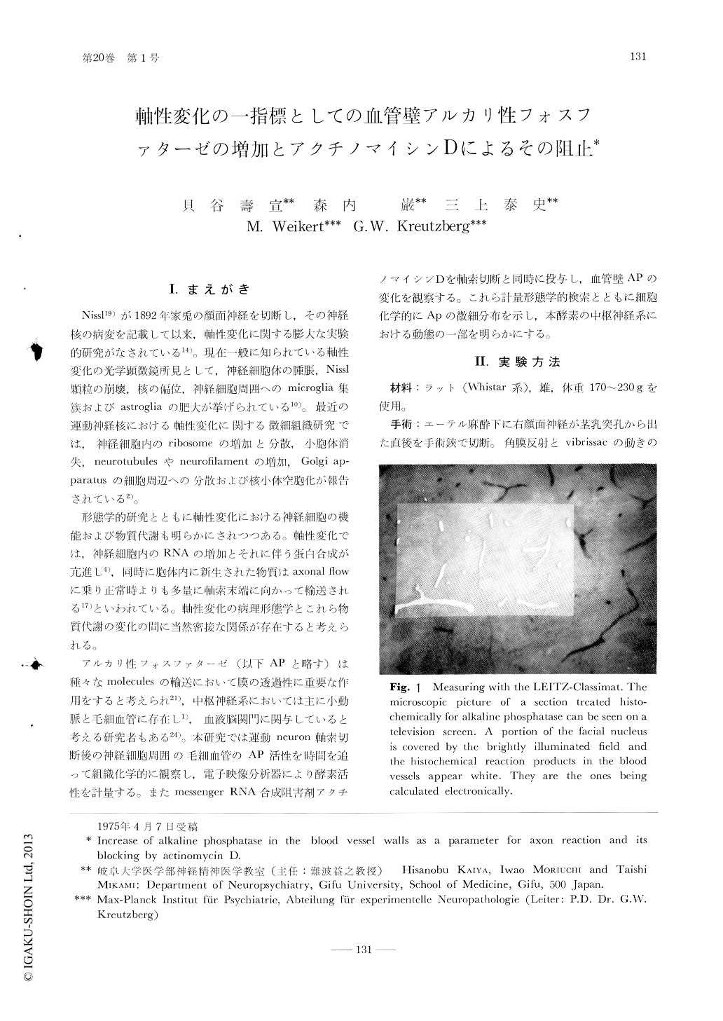
It was shown in the rat facial nucleus that increase of alkaline phosphatase activity in the blood vessels is a good parameter of the axon reaction. LEITZ-Classimat picture analyser demonstrated that this enzymatic activity begins to increase in the blood vessels 12 hours after the axon transection. The enzymatic activity obtained its peak 5 days after the operation. No difference of enzymatic activity between the nucleus undergoing axon reaction 13 days and the control site was recognized. Actinomycin D, messenger RNA synthesis inhibitor, could block the increase of alkaline phosphatase activity in the blood vessels.

Copyright © 1976, Igaku-Shoin Ltd. All rights reserved.


