Japanese
English
- 有料閲覧
- Abstract 文献概要
- 1ページ目 Look Inside
Ⅰ.小脳の気脳断層撮影法
レントゲン的に小脳の形態を観察するのには血管撮影法と気脳撮影法とがある。前者は輪郭を線で描写するため,以下に述べるような小脳各部の容積減少を観察分析する目的には十分でない。
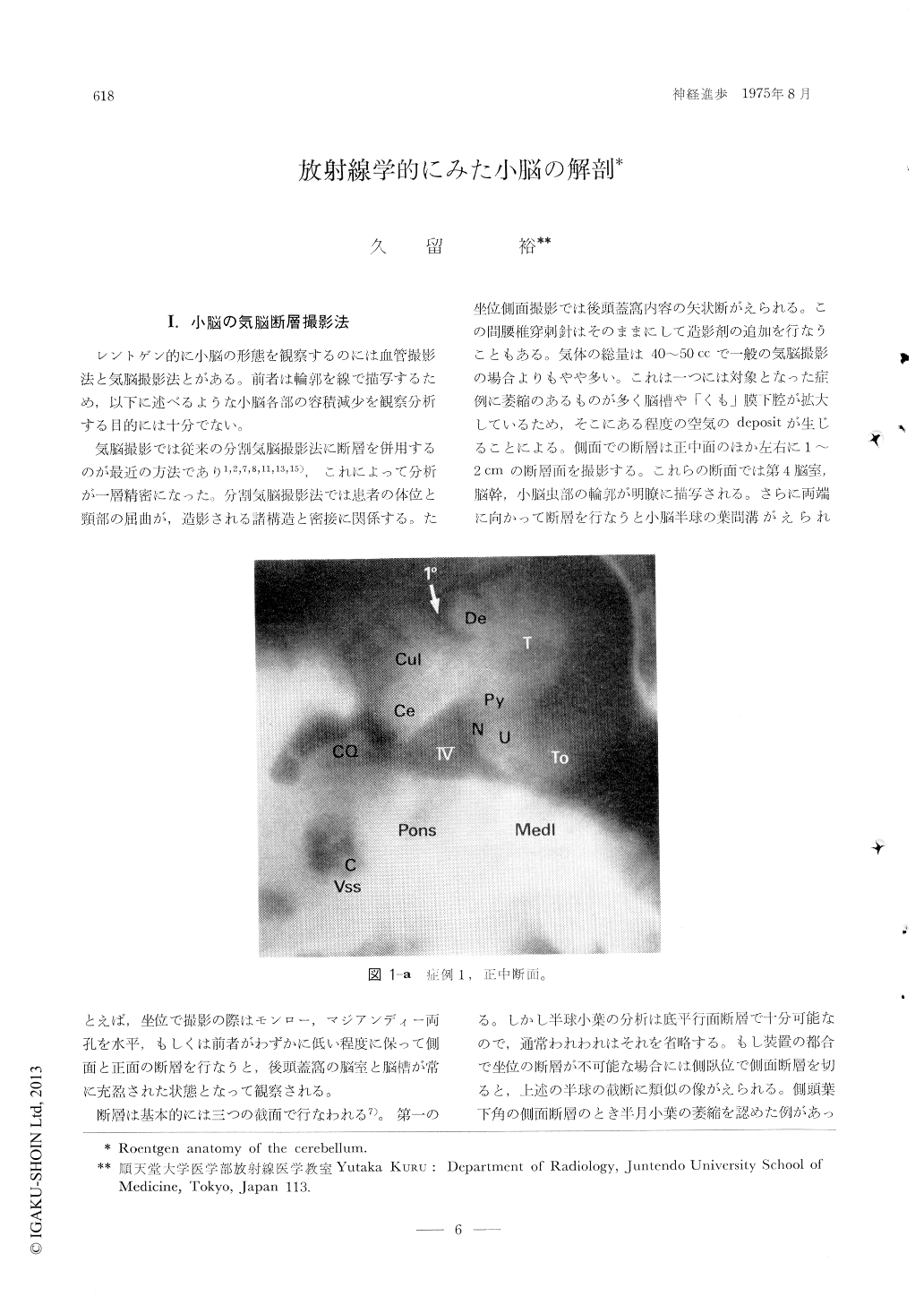
気脳撮影では従来の分割気脳撮影法に断層を併用するのが最近の方法であり1,2,7,8,11,13,15),これによって分析が一層精密になった。分割気脳撮影法では患者の体位と頸部の屈曲が,造影される諸構造と密接に関係する。たとえば,坐位で撮影の際はモンロー,マジアンディー両孔を水平,もしくは前者がわずかに低い程度に保って側面と正面の断層を行なうと,後頭蓋窩の脳室と脳槽が常に充盈された状態となって観察される。
Pneumoencephalotomography of the posterior fossa structure was used to detect a mass lesion in the fossa or a cerebellar atrophy. Both lateral and frontal tomographic cuts by a patient sitting in the erect position were established by a number of investigators. The author adds another cuts by a patient lying prone to make views of the structures at the horizontal plane. These tomo-grams have to be called "basiparallel cuts", as seen on the anatomical text, and they allow to study small macroscopic changes of the cerebellum as well as those on the fourth ventricle wall.
Copyright © 1975, Igaku-Shoin Ltd. All rights reserved.