Japanese
English
- 有料閲覧
- Abstract 文献概要
- 1ページ目 Look Inside
- サイト内被引用 Cited by
まえがき
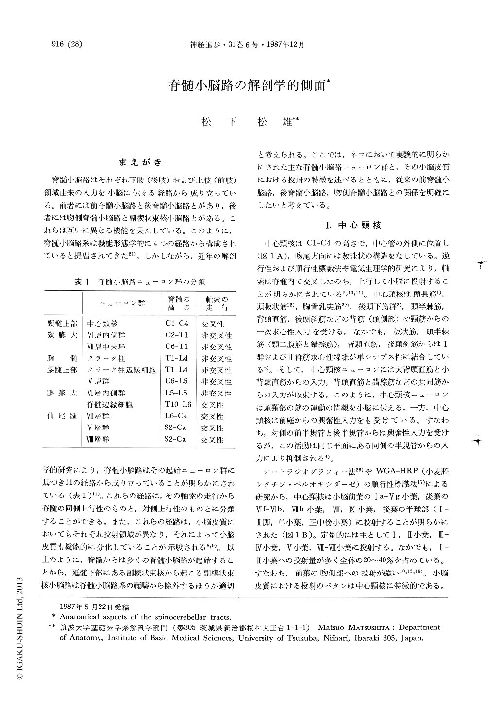
脊髄小脳路はそれぞれ下肢(後肢)および上肢(前肢)領域由来の入力を小脳に伝える経路から成り立っている。前者には前脊髄小脳路と後脊髄小脳路とがあり,後者には吻側脊髄小脳路と副楔状束核小脳路とがある。これらは互いに異なる機能を果たしている。このように,脊髄小脳路系は機能形態学的に4つの経路から構成されていると提唱されてきた21)。しかしながら,近年の解剖学的研究により,脊髄小脳路はその起始ニューロン群に基づき11の経路から成り立っていることが明らかにされている(表1)11)。これらの経路は,その軸索の走行から脊髄の同側上行性のものと,対側上行性のものとに分類することができる。また,これらの経路は,小脳皮質においてもそれぞれ投射領域が異なり,それによって小脳皮質も機能的に分化していることが示唆される8,9)。以上のように,脊髄からは多くの脊髄小脳路が起始することから,延髄下部にある副楔状束核から起こる副楔状束核小脳路は脊髄小脳路系の範疇から除外するほうが適切と考えられる。ここでは,ネコにおいて実験的に明らかにされた主な脊髄小脳路ニューロン群と,その小脳皮質における投射の特徴を述べるとともに,従来の前脊髄小脳路,後脊髄小脳路,吻側脊髄小脳路との関係を明確にしたいと考えている。
The spinocerebellar tracts originate from various groups of neurons located throughout the length of the spinal cord. They are classified into 11 tracts according to their cells of origin and their course within the spinal cord. Studies using the anterograde tracing technique reveal that each neuronal group projects to specific areas in the horizontal plane of the cerebellar lobules. Each projection area differs in the apicobasal, medio-lateral and rostrocaudal extent of the lobules.
(1) The spinocerebellar tract in the upper cervical segments originates from the central cervical nucleus, which lies lateral to the central canal at levels of the C1 to the C4 segments in the cat. The tract crosses in the spinal cord, ascends contralaterally, and projects bilaterally to all lobules of the anterior lobe, lobule VI, sublobule VIIb, lobules VIII and IX, crus I and II, the simple lobule and the paramedian lobule. The projections are most abundant in lobules I and II, accounting for 20~40% of the total quantity. The spinocerebellar tract arising from the central cervical nucleus projects to three longitudinal areas in the vermis, zones Al, Al-A2 and A2-B of Voogd. These areas are confined to the basal part in all lobules of the anterior lobe.

Copyright © 1987, Igaku-Shoin Ltd. All rights reserved.


