Japanese
English
- 有料閲覧
PPV購入案内
1,320円 (1,200円+税10%) こちらは、659ページから668ページの文献です。購入には医書.jp本会員登録が必要です。
会員登録完了後に改めて上記「購入サイトへ」を選択してください。
または「購入サイトへ」選択後に「購入ログイン」、「新規会員登録」の順に進んでください。
会員登録について詳しくはをご確認ください。
- Abstract 文献概要
- 1ページ目 Look Inside
I.はじめに
Computerized Axial Tomography(以下CATと略する)の出現によって脳の形態的診断は画期的な進歩を遂げたといっても過言ではない。
CATでは脳室はもちろんのこといくつかの脳槽も,脳実質の物質構成の差も画像として描写される。従来の神経放射線学的検査のうち,このCATの登場によって最も検査件数の減少したのが気脳撮影法であるといわれる1)。はたして今日の時点で気脳撮影法は不必要な検査となったのであろうか。
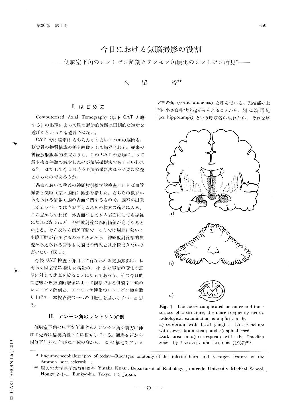
Since establishement of the CAT scanner (1973), pneumoencephalographic examinations have remarkably decreased in practice of each clinic. If a small change sited close to or within the ventricle has to be examined, this is still a target of refined techniques of pneumoencephalography. YAKOVLEV and LECOURS by their inspection of the myelinization cycle recognized that the central nervous system can be divided into three zones and that the "median zone" includes median thalamus, hypothalamus, hippocampal rudiment, septal area and hippocampus.
Copyright © 1976, Igaku-Shoin Ltd. All rights reserved.