Japanese
English
- 有料閲覧
- Abstract 文献概要
- 1ページ目 Look Inside
I.はじめに
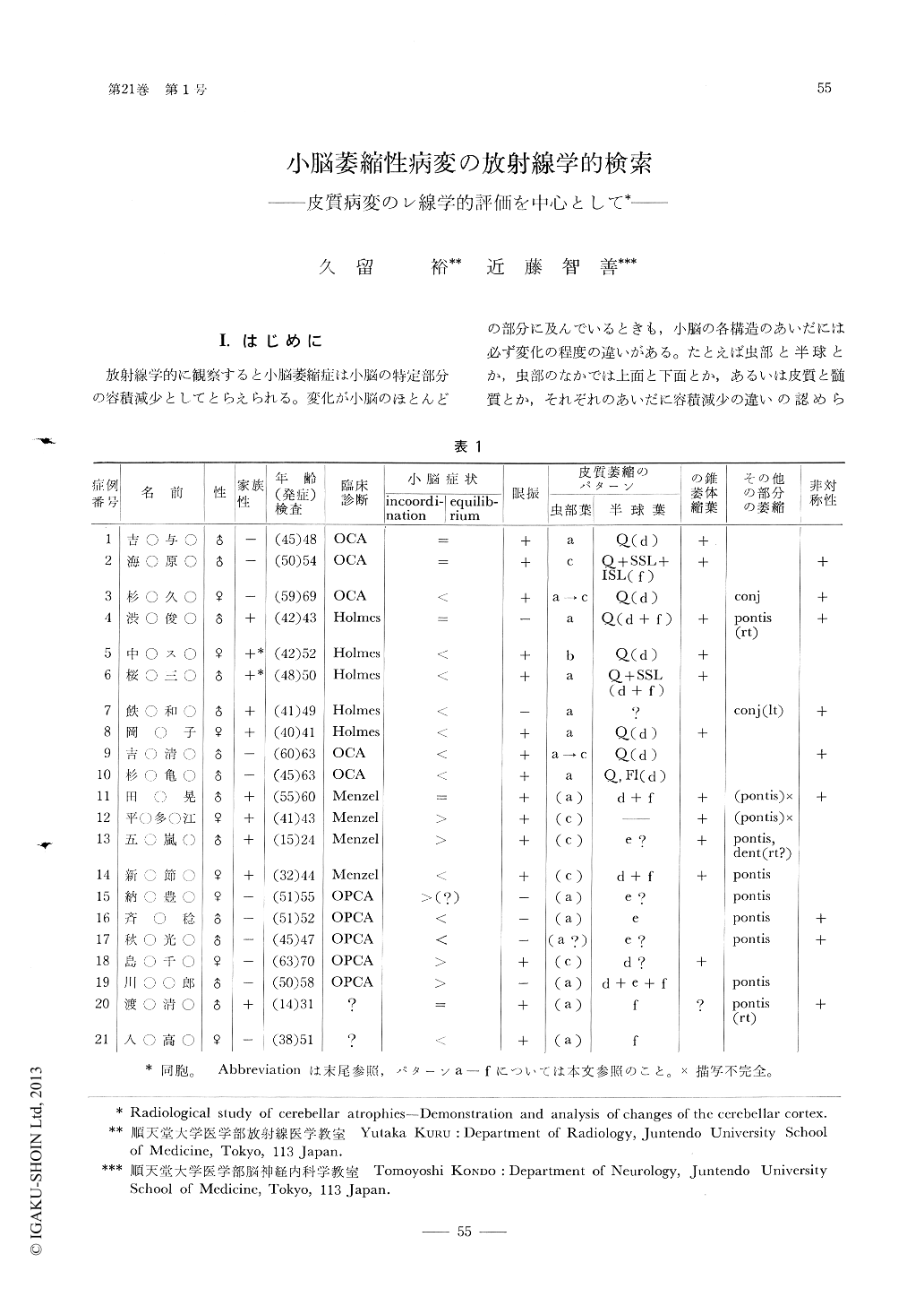
放射線学的に観察すると小脳萎縮症は小脳の特定部分の容積減少としてとらえられる。変化が小脳のほとんどの部分に及んでいるときも,小脳の各構造のあいだには必ず変化の程度の違いがある。たとえば虫部と半球とか,虫部のなかでは上面と下面とか,あるいは皮質と髄質とか,それぞれのあいだに容積減少の違いの認められるのが普通である。これらのパターン,またはパターンの組合わせから気脳撮影の所見のみで特定の小脳萎縮症と診断できることも稀ではない。しかし一般的にいうと,同じようなパターンを示すいくつかの疾患があって,気脳撮影の所見は他の臨床所見と比較検討されて最終的な診断を下さなければならない場合の方がむしろはるかに多い。
小脳萎縮に関する気脳像の報告のなかで,サルペトリエールの82例を取り扱ったBories,Fredy et Rosier3)の研究がもっとも秀れた,総括的なものであった。ただ彼らは半球の変化を充分描写しうる方法をもっていなかったため,半球皮質に病変の主座のあるべき疾患のレ線上の鑑別が必ずしも完全でなく,症例のtabulationでは虫部に主病変を示す例が不釣合に多くなってしまっている。
As we have reported previously, the three-directional tomography by air study is able to demonstrate nearly all the cortex of the cerebellum and this makes it possible to find a small macroscopic change of the structure. Briefly speaking, the sagittal cuts show the vermis, the frontal, the brainstem with the cerebellar peduncles, and the basiparallel, the cerebellar hemispheres. Some components of the fourth ventricle walls are also visualized on these tomograms. In these views various types of a cerebellar atrophy can be analyzed as for location and extent of the loss of the cortical elements.
Copyright © 1977, Igaku-Shoin Ltd. All rights reserved.