Japanese
English
- 有料閲覧
- Abstract 文献概要
- 1ページ目 Look Inside
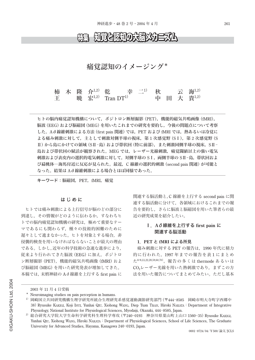
ヒトの脳内痛覚認知機構について,ポジトロン断層撮影(PET),機能的磁気共鳴画像(fMRI),脳波(EEG)および脳磁図(MEG)を用いたこれまでの研究を要約し,今後の問題点について考察した。Aδ線維刺激による方法(first pain関連)では,PETおよびfMRIでは,熱あるいは冷覚による痛み刺激に対して,主として刺激対側半球の視床,第1次感覚野(SⅠ),第2次感覚野(SⅡ)から島にかけての領域(SⅡ-島)および帯状回(特に前部),また刺激同側半球の視床,SⅡ-島および帯状回の賦活が観察された。MEGでは,レーザー光線刺激,痛覚閾値以上の強い電気刺激および表皮内の選択的電気刺激に対して,対側半球のSⅠ,両側半球のSⅡ-島,帯状回および扁桃体―海馬付近に反応が見られた。最近,C線維の選択的刺激(second pain関連)が可能となった。結果はAδ線維刺激による場合とほぼ同様であった。
We reviewed the recent progress in the ability of neuroimaging studies such as positron emission tomography(PET), functional magnetic resonance imaging(fMRI), electroencephalography(EEG)and magnetoencephalography(MEG)to detect pain perception in humans. PET and fMRI, which reflect metabolic or blood flow changes by painful stimulation, have excellent spatial resolution in order of mm, but their temporal resolution is not very high. In contrast, EEG and MEG, which reflect physiological changes, have an excellent temporal resolution in order of msec, however, it is difficult for EEG and MEG to detect activities in the deep areas such as the thalamus.
For recording activities following Aδfiber stimulation relating to the first pain,PET and fMRI are usually recorded using thermode or CO2laser stimulation as the painful heat stimuli. Although the results reported from several institutes have not been completely the same, significant increases of activities were found in the thalamus, insula, anterior cingulate cortex and the secondary somato sensory cortex(SⅡ)in the hemisphere contralateral to the stimulation, and the thalamus, insula and anterior cingulate cortex in the ipsilateral hemisphere in most reports. In addition, significant increases of activity were reported in the supplementary motor area(SMA), premotor cortex, lenticular nucleus and the cerebellum in the contralateral hemisphere, and in the premotor cortex, putamen and the cerebellum in the ipsilateral hemisphere, as well as the cerebellar vermis and midbrain in some institutes. Results following painful cold pain or intramuscular pain were not fundamentally different from results following heat stimulation. The role of the primary somato sensory cortex(SⅠ)in the contralateral hemisphere is still controversial, but recent studies have confirmed activities there, though it was rather small.
Since the spatial resolution of EEG is not very high in order of cm, MEG is useful to detect activated areas following painful stimulation. MEG on pain perception were usually recorded following painful CO2 laser stimulation, but our new method, epidermal stimulation(ES), is also very useful. The primary small activity was recorded from the SⅠ(probably in area 1)in the hemisphere contralateral to the stimulation. Then, SⅡand insula were activated with the second activity in SⅠ. These three regions were activated in parallel with almost the same time period. This is a very characteristic finding in pain perception. Then, the cingulate cortex and medial temporal area(MT)around the amygdala and hippocampus were activated. In the hemisphere ipsilateral to the stimulation as well, the above regions were activated, except for SⅠ. Therefore, SⅠis considered to play a main role in localization of the stimulus point,the SⅡand insula are important sites for pain perception, and the cingulate and MT are mainly responsible for cognitive or emotional aspects for pain perception.
For recording activities following C fiber stimulation relating to the first pain, intradermal injection of capsaicin was used for recording PET and fMRI. Similar regions to those in Aδfiber stimulation study were activated, but the results were not consistent among the studies. For recording EEG and MEG, we recently developed new method, that is, applying weaker CO2 laser stimuli to tiny areas of the skin. MEG findings following C fiber stimulation were also similar to those following Aδfiber stimulation. However, the effect of sleep and attention on MEG following C fiber stimulation was much larger than that following Aδfiber stimulation. This finding may suggest greater effect of cognitive or emotional functions on second pain than the first pain.
(Received:October 17, 2003)

Copyright © 2004, Igaku-Shoin Ltd. All rights reserved.


