Japanese
English
- 有料閲覧
PPV購入案内
1,320円 (1,200円+税10%) こちらは、396ページから405ページの文献です。購入には医書.jp本会員登録が必要です。
会員登録完了後に改めて上記「購入サイトへ」を選択してください。
または「購入サイトへ」選択後に「購入ログイン」、「新規会員登録」の順に進んでください。
会員登録について詳しくはをご確認ください。
- Abstract 文献概要
- 1ページ目 Look Inside
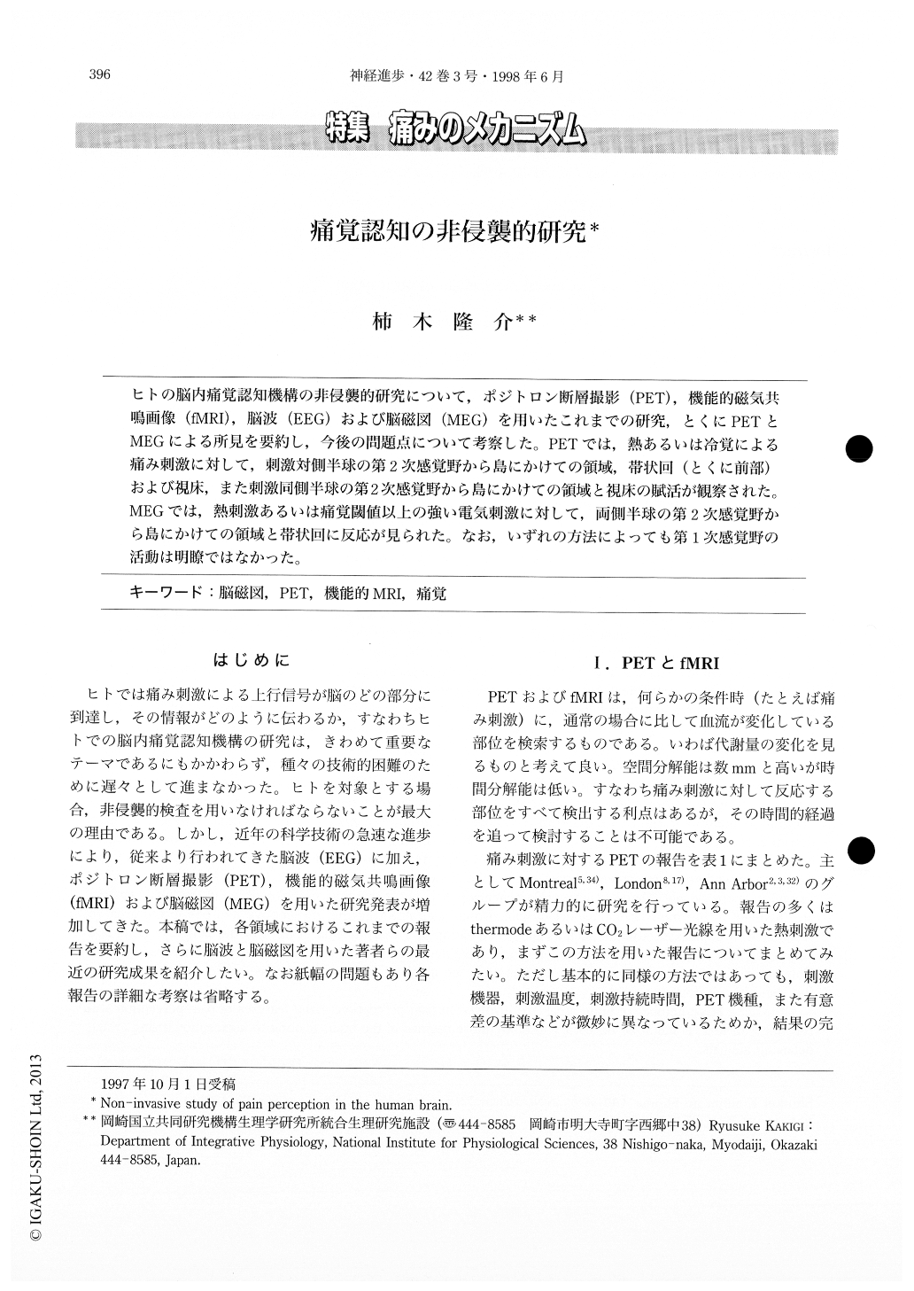
ヒトの脳内痛覚認知機構の非侵襲的研究について,ポジトロン断層撮影(PET),機能的磁気共鳴画像(fMRI),脳波(EEG)および脳磁図(MEG)を用いたこれまでの研究,とくにPETとMEGによる所見を要約し,今後の問題点について考察した。PETでは,熱あるいは冷覚による痛み刺激に対して,刺激対側半球の第2次感覚野から島にかけての領域,帯状回(とくに前部)および視床,また刺激同側半球の第2次感覚野から島にかけての領域と視床の賦活が観察された。MEGでは,熱刺激あるいは痛覚閾値以上の強い電気刺激に対して,両側半球の第2次感覚野から島にかけての領域と帯状回に反応が見られた。なお,いずれの方法によっても第1次感覚野の活動は明瞭ではなかった。
Non-invasive studies of pain perception in the human brain have been performed by positron emission tomography (PET), functional magnetic resonance imaging (fMRI) , electroencephalography (EEG) and magnetoencephalography (MEG). PET and fMRI, which reflect metabolic changes by painful stimulation, have excellent spatial resolution in the order of mm, but their temporal resolution is not so high. In contrast, EEG and MEG, which reflect physiological changes, have excellent temporal resolution in the order of msec. However, it is difficult for EEG and MEG to detect activities in the deep areas such as the thalamus.
Copyright © 1998, Igaku-Shoin Ltd. All rights reserved.