Japanese
English
- 有料閲覧
- Abstract 文献概要
- 1ページ目 Look Inside
患者 62歳,男性.
主訴 なし.
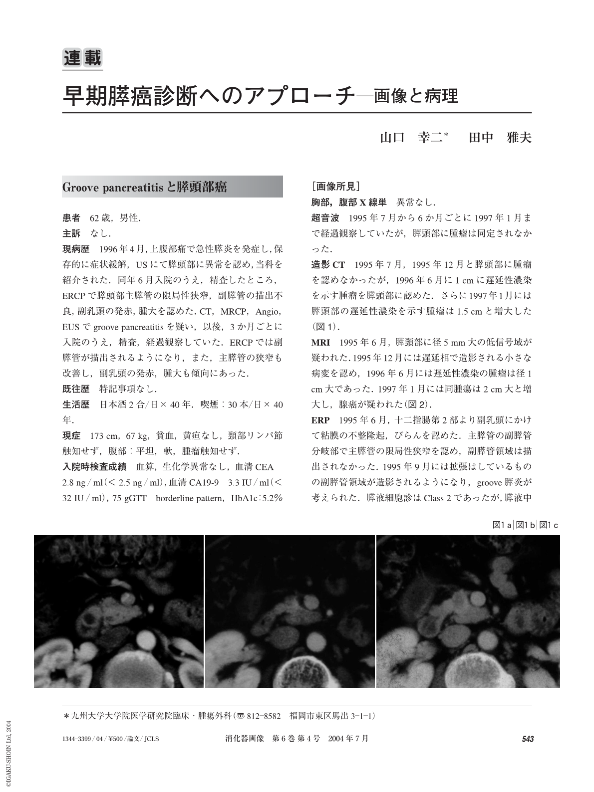
現病歴 1996年4月,上腹部痛で急性膵炎を発症し,保存的に症状緩解,USにて膵頭部に異常を認め,当科を紹介された.同年6月入院のうえ,精査したところ,ERCPで膵頭部主膵管の限局性狭窄,副膵管の描出不良,副乳頭の発赤,腫大を認めた.CT,MRCP,Angio,EUSでgroove pancreatitisを疑い,以後,3か月ごとに入院のうえ,精査,経過観察していた.ERCPでは副膵管が描出されるようになり,また,主膵管の狭窄も改善し,副乳頭の発赤,腫大も傾向にあった.
A 62-year-old man developed acute pancreatitis and was followed up under the diagnosis of groove pancreatitis. Follow-up imagings were taken every three months. One year after, a pancreatic mass with delayed enhancement was noticed on CT. MRI showed also a pancreatic head mass with delayed enhancement, measuring 1 cm. ERP showed a stenosis of the main pancreatic duct. EUS demonstrated a well-defined hypoechoic pancreatic head mass. Pylorus preserving pancreatoduodenectomy with D2 lymph node dissection was performed under the diagnosis of pancreatic head cancer. Intraoperative radiation therapy(18 Gy)was also done. Histological examination showed a well differentiated adenocarcinoma, measuring 1.5 cm and groove pancreatitis. Lymph node metastasis was not evident and surgical margins were free of cancer cells. He died of local recurrence and lung metastasis 5 years and 3 months after operation.
(Shokakigazo 2004 ; 6 : 543―547)

Copyright © 2004, Igaku-Shoin Ltd. All rights reserved.


