Japanese
English
- 有料閲覧
- Abstract 文献概要
- 1ページ目 Look Inside
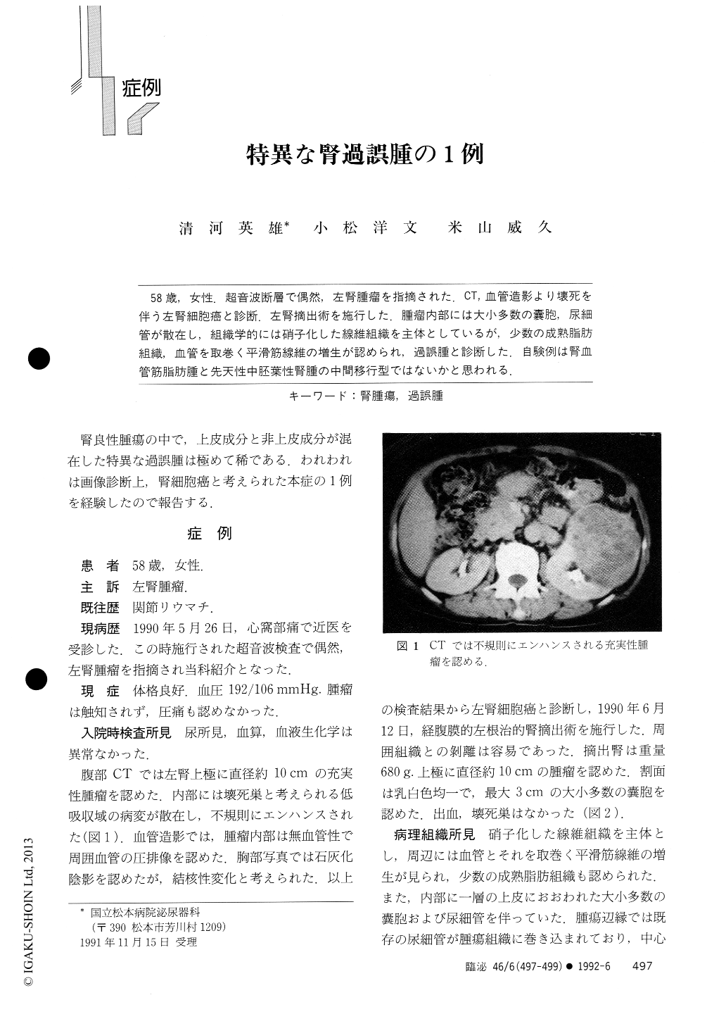
58歳,女性.超音波断層で偶然,左腎腫瘤を指摘された.CT.血管造影より壊死を伴う左腎細胞癌と診断.左腎摘出術を施行した.腫瘤内部には大小多数の嚢胞,尿細管が散在し,組織学的には硝子化した線維組織を主体としているが,少数の成熟脂肪組織血管を取巻く平滑筋線維の増生が認められ,過誤腫と診断した.自験例は腎血管筋脂肪腫と先天性中胚葉性腎腫の中間移行型ではないかと思われる.
A 58-year-old woman was incidentally found to have a left renal mass on ultrasonography. CT showed a solid, irregularly enhanced mass of 10 cm in diameter in the left kidney. The mass was avascular on angiography. The patient underwent nephrectomy. The mass was pale yellow. Histologically it consisted of hyalinized fibrous tissue intermingled with cystic and tubular structures and mature fat tissue, and vessels surrounded by smooth muscle. The diagnosis of renal hamartoma was made. This tumor is considered to be an intermediate form between angiomyolipoma and congenital mesoblastic nephroma.

Copyright © 1992, Igaku-Shoin Ltd. All rights reserved.


