Japanese
English
- 有料閲覧
- Abstract 文献概要
- 1ページ目 Look Inside
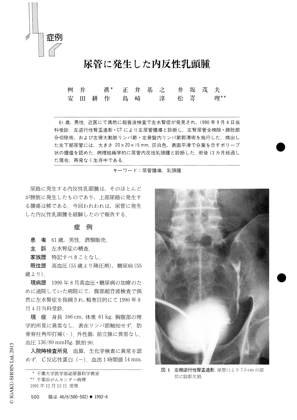
61歳,男性.近医にて偶然に超音波検査で左水腎症が発見され,1990年9月4日当科受診.左逆行性腎盂造影・CTにより左尿管腫瘍と診断し,左腎尿管全摘除.膀胱部分切除術,および左傍大動脈リンパ節・左骨盤内リンパ節郭清術を施行した.摘出した左下部尿管には,大きさ20×20×15mm,灰白色,表面平滑で分葉を示すポリープ状の腫瘤を認めた.病理組織学的に尿管内反性乳頭腫と診断した.術後13ヵ月経過した現在,再発なく生存中である.
A 61-years old man was admitted because of incidentally discovered left hydronephrosis by abdominal ultrasonography. Physical examination was normal and laboratory data were all within normal limits. No malignant cells were found on urine cytology. Intravenous urography demonstrated a normal right kidney and a nonfunctioning left kidney. Computed tomography (CT) revealed a left hydronephrosis and a dilatation of middle and upper ureter. Left retrograde and antegrade pyeloureterography revealed a sharp-edged filling defect in the left lower ureter which was diagnoed as ureteral tumor.

Copyright © 1992, Igaku-Shoin Ltd. All rights reserved.


