Japanese
English
- 有料閲覧
- Abstract 文献概要
- 1ページ目 Look Inside
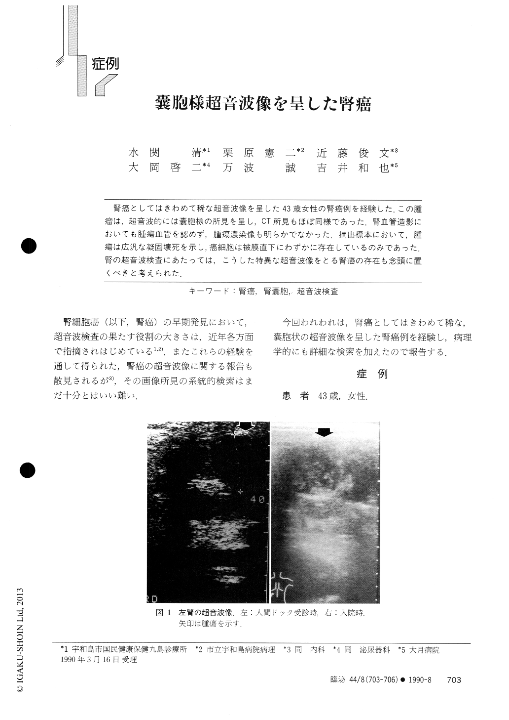
腎癌としてはきわめて稀な超音波像を呈した43歳女性の腎癌例を経験した.この腫瘤は,超音波的には嚢胞様の所見を呈し,CT所見もほぼ同様であった.腎血管造影においても腫瘍血管を認めず,腫瘍濃染像も明らかでなかった.摘出標本において,腫瘍は広汎な凝固壊死を示し,癌細胞は被膜直下にわずかに存在しているのみであった.腎の超音波検査にあたっては,こうした特異な超音波像をとる腎癌の存在も念頭に置くべきと考えられた,
The tumor was seen in the upper pole of left kidney in a 43-year-old woman. Ultrasonographically the tumor appeared sonolucent mass mimicking simple renal cyst. Computed tomography revealed that the tumor had an attenuation value of 14.43 Hounsefield unit, suggesting cystic lesion again. Renal angiography showed neither tumor vessels nor tumor stain. Histologic examination of the surgically removed kidney revealed that the tumor had thick capsule, and all the tumor cells had undergone coagulative necrosis except for those adjacent to the capsules.

Copyright © 1990, Igaku-Shoin Ltd. All rights reserved.


