Japanese
English
- 有料閲覧
- Abstract 文献概要
- 1ページ目 Look Inside
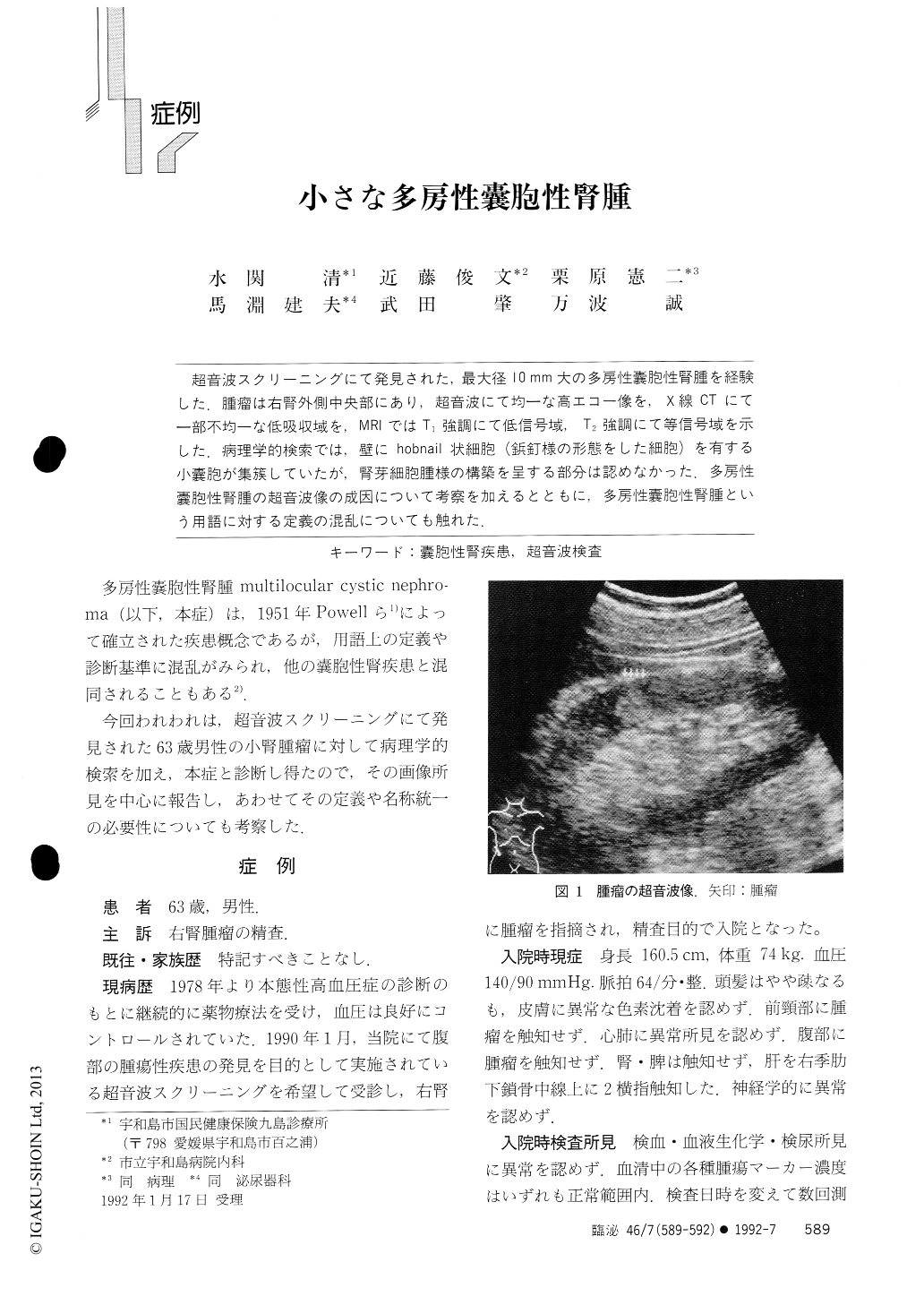
超音波スクリーニングにて発見された,最大径10mm大の多房性嚢胞性腎腫を経験した.腫瘤は右腎外側中央部にあり,超音波にて均一な高エコー像を,X線CTにて一部不均一な低吸収域を,MRIではT1強調にて低信号域,T2強調にて等信号域を示した.病理学的検索では,壁にhobnail状細胞(鋲釘様の形態をした細胞)を有する小嚢胞が集簇していたが,腎芽細胞腫様の構築を呈する部分は認めなかった.多房性嚢胞性腎腫の超音波像の成因について考察を加えるとともに,多房性嚢胞性腎腫という用語に対する定義の混乱についても触れた.
A multilocular cystic nephroma, measuring l0 mm in the greatest diameter, occurred in a 63-year-o1dJapanese male without any significant clinical symptoms. The tumor exhibited a homogeneously hyperechoicappearance, mimicking an angiomyolipoma. Histologically, the tumor was well demarcated from surround-ing renal tissues, and consisted of multiple loculi with epithelial lining. Some of the epithelia showedcharacteristic appearances called hobnail cell pattern.

Copyright © 1992, Igaku-Shoin Ltd. All rights reserved.


