Japanese
English
- 有料閲覧
- Abstract 文献概要
- 1ページ目 Look Inside
はじめに
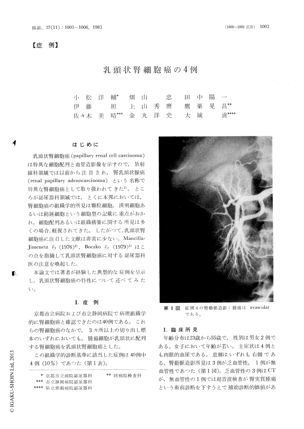
乳頭状腎細胞癌(papillary renal cell carcinoma)は特異な細胞配列と血管造影像を示すので,放射線科領域では以前から注目され,腎乳頭状腺癌(renal papillary adenocarcinoma)という名称で特異な腎細胞癌として取り扱われてきた1)。ところが泌尿器科領域では,とくに本邦においては,腎細胞癌の組織学的所見は顆粒細胞,淡明細胞あるいは紡錘細胞という細胞型の記載に重点がおかれ,細胞配列あるいは組織構築に関する所見は多くの場合,軽視されてきた。したがつて,乳頭状腎細胞癌に注目した文献は非常に少ない。Mancilla—Jimenetzら(1976)2),Boczkoら(1979)3)はこの点を指摘して乳頭状腎細胞癌に対する泌尿器科医の注意を喚起した。
本論文では著者が経験した典型的な症例を呈示し,乳頭状腎細胞癌の特性について述べてみたい。
In varying reports concerning the clinical prognosis of renal cell carcinoma, excessive attention was primarily directed to cell type while almost totally ignoring the histologic organization in Japan.
In the retrospective review of 40 renal cell carcinoma, 4 cases were found to be papillary renal cell carcinoma. Angiographic and histological studies of these cases revealed that papillary renal cell carci-noma was different from the more common form of renal cell carcinoma in several respects.

Copyright © 1983, Igaku-Shoin Ltd. All rights reserved.


