Japanese
English
- 有料閲覧
- Abstract 文献概要
- 1ページ目 Look Inside
緒言
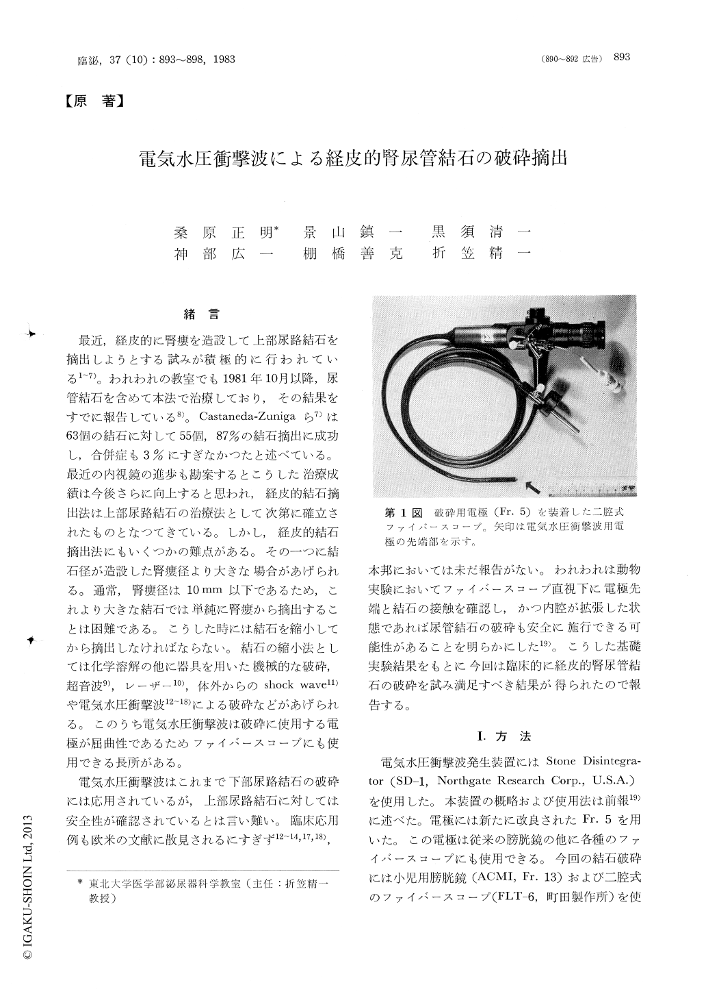
最近,経皮的に腎瘻を造設して上部尿路結石を摘出しようとする試みが積極的に行われている1〜7)。われわれの教室でも1981年10月以降,尿管結石を含めて本法で治療しており,その結果をすでに報告している8)。Castaneda-Zunigaら7)は63個の結石に対して55個,87%の結石摘出に成功し,合併症も3%にすぎなかつたと述べている。最近の内視鏡の進歩も勘案するとこうした治療成績は今後さらに向上すると思われ,経皮的結石摘出法は上部尿路結石の治療法として次第に確立されたものとなつてきている。しかし,経皮的結石摘出法にもいくつかの難点がある。その一つに結石径が造設した腎瘻径より大きな場合があげられる。通常,腎瘻径は10mm以下であるため,これより大きな結石では単純に腎瘻から摘出することは困難である。こうした時には結石を縮小してから摘出しなければならない。結石の縮小法としては化学溶解の他に器具を用いた機械的な破砕,超音波9),レーザー10),体外からのshock wave11)や電気水圧衝撃波12〜18)による破砕などがあげられる。このうち電気水圧衝撃波は破砕に使用する電極が屈曲性であるためファイパースコープにも使用できる長所がある。
Percutaneous electrohydraulic nephro-ureterolithotripsy using a 13 Fr. cystoscope or a double lumi-nal fiberscope (Fr. 16) was successfully performed on seven patients (four pyclolithiasis and three ureterolithiasis). The disintegration was performed via an old nephrostomy or a newly constructed percutaneous nephrostomy under ultrasonic guidance. The size of disintegrated calculi was more than ten mm in minimal diameter and the composition was magnesium ammonium phosphate in one, calcium oxalate in three and mixed calcium oxalate and calcium phosphate in three patients.

Copyright © 1983, Igaku-Shoin Ltd. All rights reserved.


