Japanese
English
- 有料閲覧
- Abstract 文献概要
- 1ページ目 Look Inside
緒言
1971年,Subramanian1)により99mTC燐酸化合物を用いる骨シンチグラフィー(以下骨シンチと略す)が開発されて以来,従来の骨親和性核種を用いる方法に比し,画像,検査時間など種々の点ですぐれていることから,本法は各種骨病変のルーチン検査法として急速に普及した。当然,これはまた前立腺癌骨転移の早期発見,治療効果の判定などにも広く用いられるようになつたが,骨X線所見との対比において画像の判定上問題があることも少なくない。これらの点に鑑み,今回著者らは過去6年間に経験した前立腺癌症例に対する骨シンチ像を臨床的に検討した。
Eighty radioisotopic bone scintiscannings were carried out on 47 patients with prostatic carcinoma experienced for the past 6 years. Results were as follows:
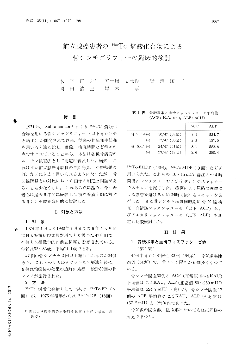
1) Skeletal abnormal uptakes (positive bone scan) were observed in 30 of 47 cases (64%), while osteoblastic and osteolytic changes of bone X-ray (positive bone survey) were noted in 24 of 47 cases (51%).
2) In 30 positive bone scan cases, mean values of ACP and ALP were 7.4 K.A. and 524.7 mIU, respectively. Both ACP and ALP showed abnormally high values.
Copyright © 1981, Igaku-Shoin Ltd. All rights reserved.