Japanese
English
- 有料閲覧
- Abstract 文献概要
- 1ページ目 Look Inside
はじめに
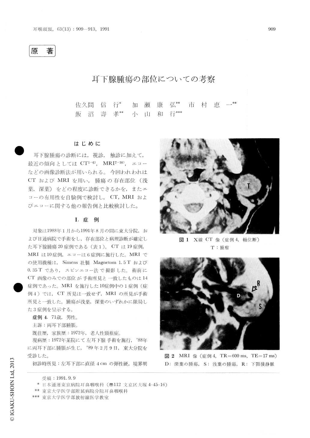
耳下腺腫瘍の診断には,視診,触診に加えて,最近の傾向としてはCT1-6),MRI7-10),エコーなどの画像診断法が用いられる。今回われわれはCTおよびMRIを用い,腫瘍の存在部位(浅葉,深葉)をどの程度に診断できるかを,またエコーの有用性を自験例で検討し,CT,MRIおよびエコーに関する他の報告例と比較検討した。
Twenty cases of parotid tumors were reported with special reference to the imging modalities. The present-day imaging modalities include CT, MRI and ultrasonography. Although CT well de-fines the tumor itself, its location is not specifi-cally determined, i.e., whether is the superficial or deep lobes. On T1-weighted images of MRI, the facial nerve appeared as a curvilinear structure of relatively low signal intensity wlthin the fatty, high-signal parotid parenchyma. Although this does not appear constantly, the identification of the facial nerve wihin the gland will definitely point the location. Ultrasonography is rather suited in differentiating between benign and malgnantlesions.

Copyright © 1991, Igaku-Shoin Ltd. All rights reserved.


