Japanese
English
原著
耳下腺単形腺腫4症例の画像診断—多形腺腫との画像上の比較
CT and MRI Findings of Monomorphic Adenoma:Report of Four Cases
田中 利善
1
,
石尾 健一郎
1
,
飯沼 壽孝
1
,
佐久間 信行
2
,
小山 和行
3
Toshiyoshi Tanaka
1
1東京大学医学部附属病院分院耳鼻咽喉科
2日本通運東京病院耳鼻咽喉科
3東京大学医科学研究所放射線科
1Department of Otolaryngology, Tokyo University Branch Hospital
pp.771-776
発行日 1992年10月20日
Published Date 1992/10/20
DOI https://doi.org/10.11477/mf.1411900607
- 有料閲覧
- Abstract 文献概要
- 1ページ目 Look Inside
はじめに
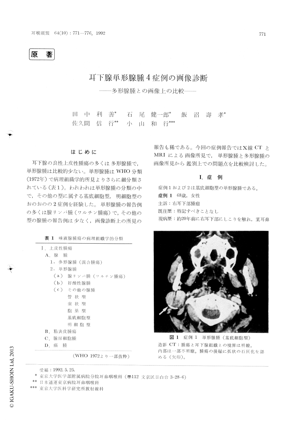
耳下腺の良性上皮性腫瘍の多くは多形腺腫で,単形腺腫は比較的少ない。単形腺腫はWHO分類(1972年)で病理組織学的所見よりさらに細分類されている(表1)。われわれは単形腺腫の分類の中で,その他の型に属する基底細胞型,明細胞型のおのおのの2症例を経験した。単形腺腫の報告例の多くは腺リンパ腫(ワルチン腫瘍)で,その他の型の腺腫の報告例は少なく,画像診断上の所見の報告も稀である。今回の症例報告ではX線CTとMRIによる画像所見で,単形腺腫と多形腺腫の画像所見から鑑別上での問題点を比較検討した。
In two cases of basal cell adenoma and two cases of clear cell adenoma the findings of CT and MRI were described, and the results were compared with those of pleomorphic adenoma.
No substantial differences were seen between monomorphic and pleomorphic adenomas in CT, MRI and RI finding.

Copyright © 1992, Igaku-Shoin Ltd. All rights reserved.


