Japanese
English
- 有料閲覧
- Abstract 文献概要
- 1ページ目 Look Inside
I.はじめに
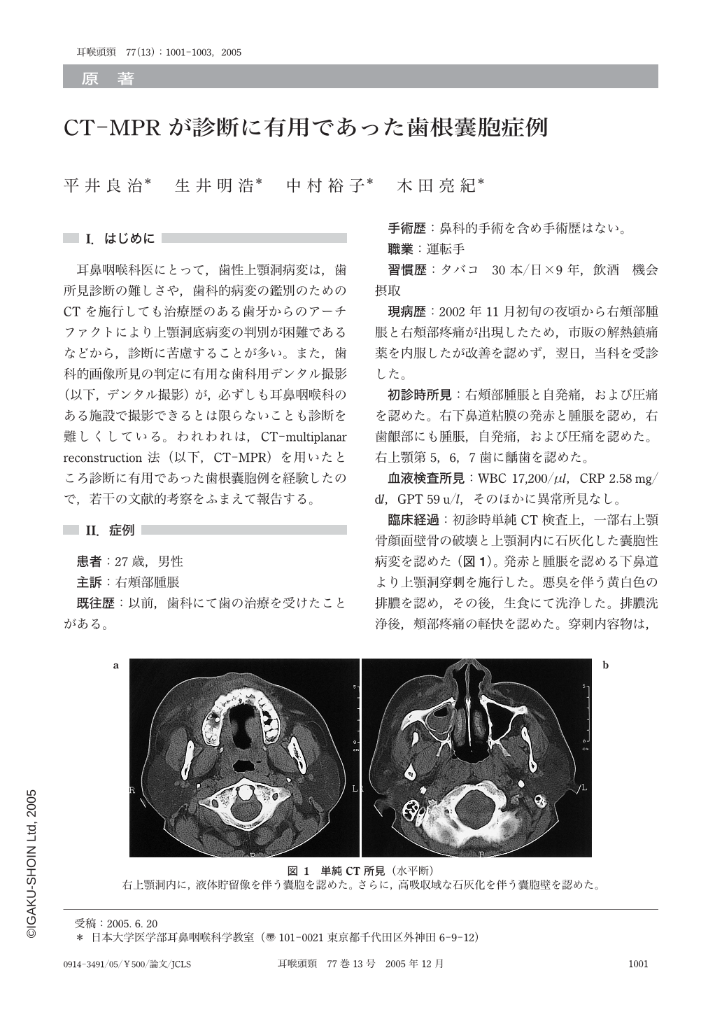
耳鼻咽喉科医にとって,歯性上顎洞病変は,歯所見診断の難しさや,歯科的病変の鑑別のためのCTを施行しても治療歴のある歯牙からのアーチファクトにより上顎洞底病変の判別が困難であるなどから,診断に苦慮することが多い。また,歯科的画像所見の判定に有用な歯科用デンタル撮影(以下,デンタル撮影)が,必ずしも耳鼻咽喉科のある施設で撮影できるとは限らないことも診断を難しくしている。われわれは,CT-multiplanar reconstruction法(以下,CT-MPR)を用いたところ診断に有用であった歯根囊胞例を経験したので,若干の文献的考察をふまえて報告する。
For otorhinolayngologists it is sometimes difficult to assess odontogenic maxillary sinus region by dental x-ray or CT. Furthermore,CT imaging causes artifacts due to dental prostheses. A 27-year-old male presented with a radicular cyst occupying the right maxillary sinus. CT with MPR revealed a radicular cyst without artifacts. For otorhinolayngologists CT with MPR is excellent modality to make a diagnosis of radicular cyst.

Copyright © 2005, Igaku-Shoin Ltd. All rights reserved.


