Japanese
English
- 有料閲覧
- Abstract 文献概要
- 1ページ目 Look Inside
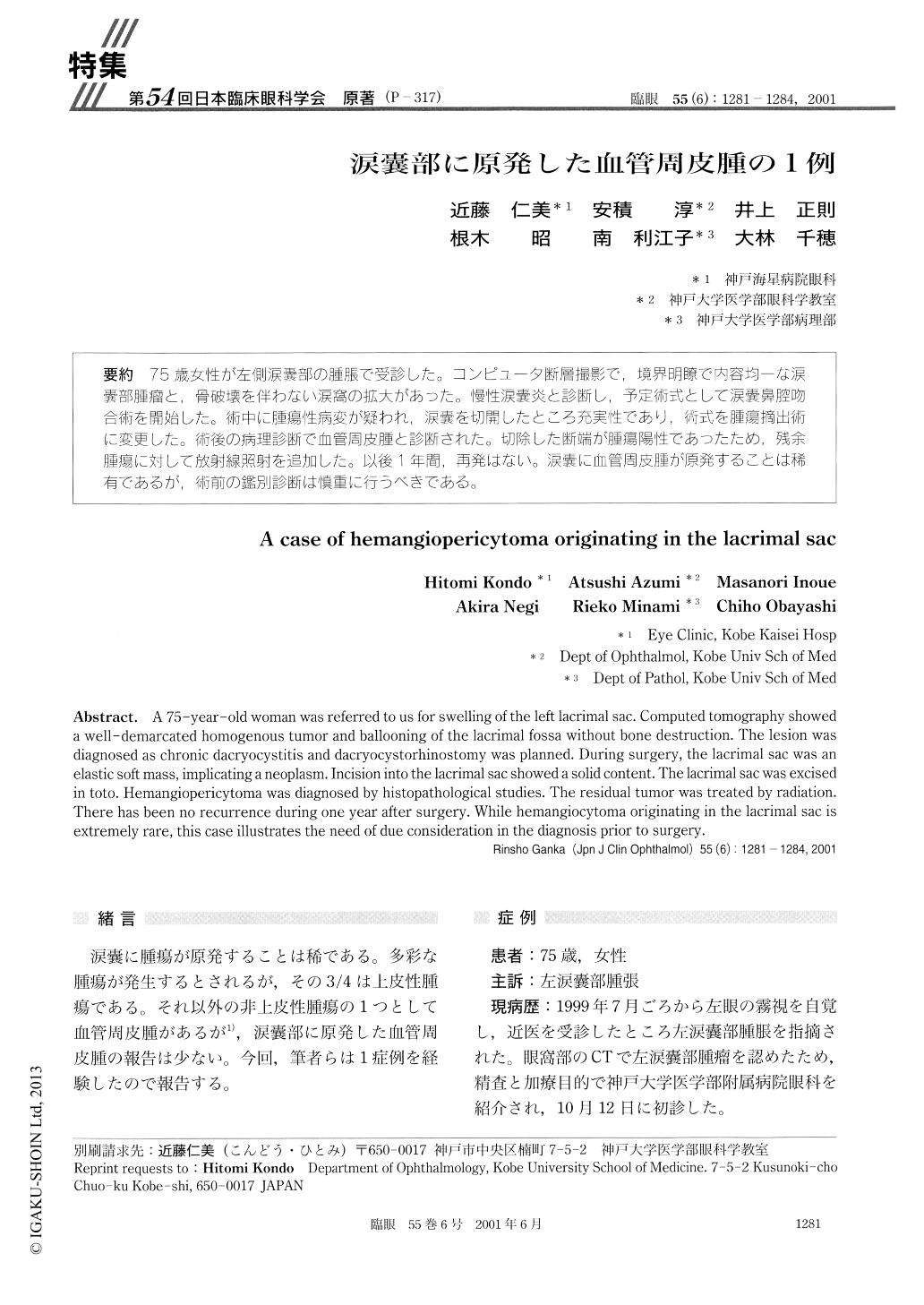
75歳女性が左側涙嚢部の腫脹で受診した。コンピュータ断層撮影で,境界明瞭で内容均一な涙嚢部腫瘤と,骨破壊を伴わない涙窩の拡大があった。慢性涙嚢炎と診断し,予定術式として涙嚢鼻腔吻合術を開始した。術中に腫瘍性病変が疑われ,涙嚢を切開したところ充実性であり,術式を腫瘍摘出術に変更した。術後の病理診断で血管周皮腫と診断された。切除した断端が腫瘍陽性であったため,残余腫瘍に対して放射線照射を追加した。以後1年間,再発はない。涙嚢に血管周皮腫が原発することは稀有であるが,術前の鑑別診断は慎重に行うべきである。
A 75-year-old woman was referred to us for swelling of the left lacrimal sac. Computed tomography showed a well-demarcated homogenous tumor and ballooning of the lacrimal fossa without bone destruction. The lesion was diagnosed as chronic dacryocystitis and dacryocystorhinostomy was planned. During surgery, the lacrimal sac was an elastic soft mass, implicating a neoplasm. Incision into the lacrimal sac showed a solid content. The lacrimal sac was excised in toto. Hemangiopericytoma was diagnosed by histopathological studies. The residual tumor was treated by radiation. There has been no recurrence during one year after surgery. While hemangiocytoma originating in the lacrimal sac is extremely rare, this case illustrates the need of due consideration in the diagnosis prior to surgery.

Copyright © 2001, Igaku-Shoin Ltd. All rights reserved.


