Japanese
English
- 有料閲覧
- Abstract 文献概要
- 1ページ目 Look Inside
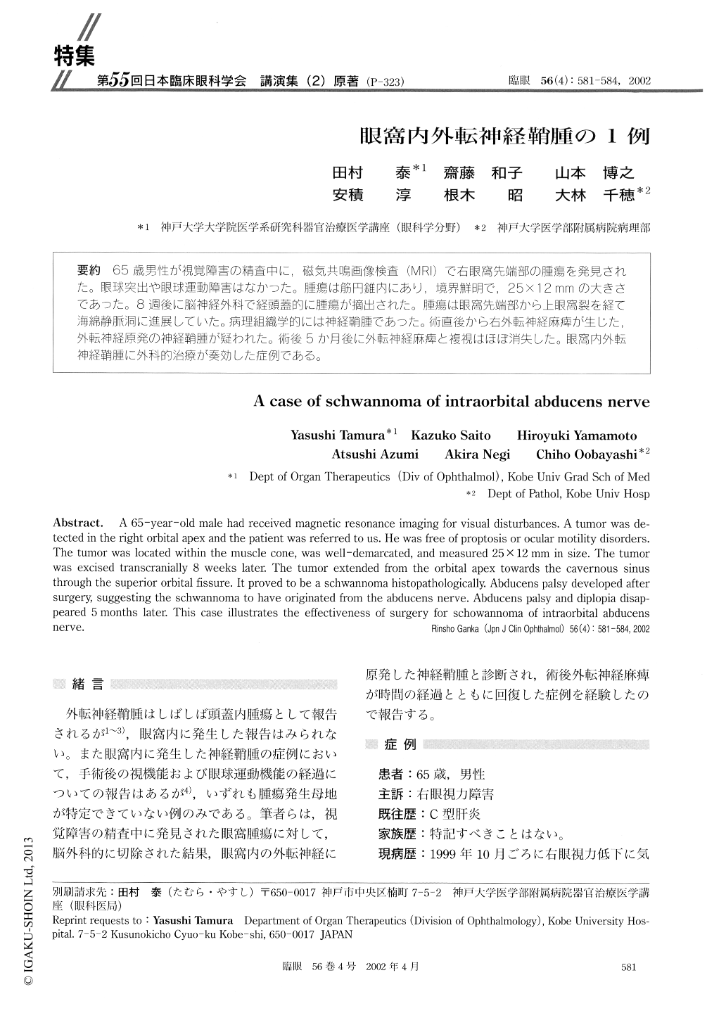
65歳男性が視覚障害の精査中に,磁気共鳴画像検査(MRDで右眼窩先端部の腫瘍を発見された。眼球突出や眼球運動障害はなかった。腫瘍は筋円錐内にあり,境界鮮明で,25×12mmの大きさであった。8週後に脳神経外科で経頭蓋的に腫瘍が摘出された。腫瘍は眼窩先端部から上眼窩裂を経て海綿静脈洞に進展していた。病理組織学的には神経鞘腫であった。術直後から右外転神経麻痺が生じた,外転神経原発の神経鞘腫が疑われた。術後5か月後に外転神経麻痺と複視はほぼ消失した。眼窩内外転神経鞘腫に外科的治療が奏効した症例である。
A 65-year-old male had received magnetic resonance imaging for visual disturbances. A tumor was de-tected in the right orbital apex and the patient was referred to us. He was free of proptosis or ocular motility disorders. The tumor was located within the muscle cone, was well-demarcated, and measured 25×12 mm in size. The tumor was excised transcranially 8 weeks later. The tumor extended from the orbital apex towards the cavernous sinus through the superior orbital fissure. It proved to be a schwannoma histopathologically. Abducens palsy developed after surgery, suggesting the schwannoma to have originated from the abducens nerve. Abducens palsy and diplopia disap-peared 5 months later. This case illustrates the effectiveness of surgery for schowannoma of intraorbital abducens nerve.

Copyright © 2002, Igaku-Shoin Ltd. All rights reserved.


