Japanese
English
- 有料閲覧
- Abstract 文献概要
- 1ページ目 Look Inside
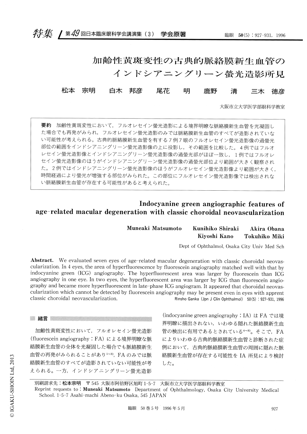
加齢性黄斑変性において,フルオレセイン螢光造影による境界明瞭な脈絡膜新生血管を光凝固した場合でも再発がみられ,フルオレセイン螢光造影のみでは脈絡膜新生血管のすべてが造影されていない可能性が考えられる。古典的脈絡膜新生血管を有する7例7眼のフルオレセイン螢光造影像の過螢光部位の範囲をインドシアニングリーン螢光造影像の上に投影し,その範囲を比較した。4例ではフルオレセイン螢光造影像とインドシアニングリーン螢光造影像の過螢光部がほぼ一致し,1例ではフルオレセイン螢光造影像のほうがインドシアニングリーン螢光造影像の過螢光部位より範囲が大きく観察された。2例ではインドシアニングリーン螢光造影像のほうがフルオレセイン螢光造影像より範囲が大きく,時間経過により螢光が増強する部位がみられた。この部位にフルオレセイン螢光造影像では検出されない脈絡膜新生血管が存在する可能性があると考えられた。
We evaluated seven eyes of age-related macular degeneration with classic choroidal neovas-cularization. In 4 eyes, the area of hyperfluorescence by fluorescein angiography matched well with that by indocyanine green (ICG) angiography. The hyperfluorescent area was larger by fluorescein than ICG angiography in one eye. In two eyes, the hyperfluorescent area was larger by ICG than fluorescein angio-graphy and became more hyperfluorescent in late-phase ICG angiogram. It appeared that choroidal neovas-cularization which cannot be detected by fluorescein angiography may be present even in eyes with apprent classic choroidal neovascularization.

Copyright © 1996, Igaku-Shoin Ltd. All rights reserved.


