Japanese
English
- 有料閲覧
- Abstract 文献概要
- 1ページ目 Look Inside
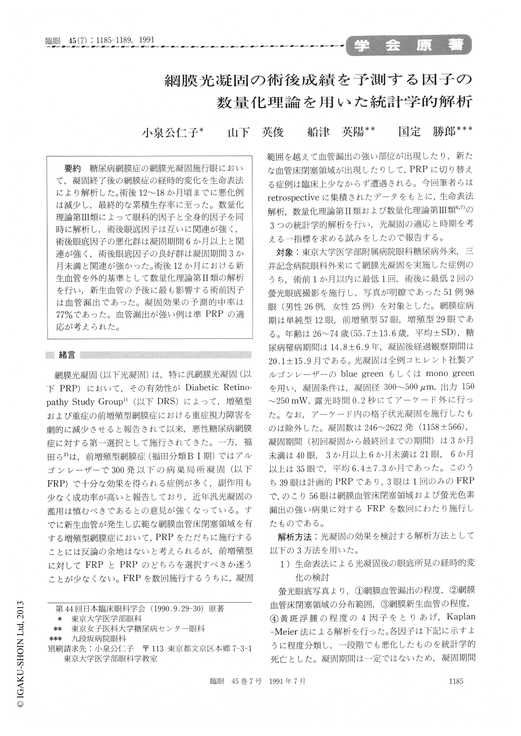
糖尿病網膜症の網膜光凝固施行眼において,凝固終了後の網膜症の経時的変化を生命表法により解析した。術後12〜18か月頃までに悪化例は減少し,最終的な累積生存率に至った。数量化理論第Ⅲ類によって眼科的因子と全身的因子を同時に解析し,術後眼底因子は互いに関連が強く,術後眼底因子の悪化群は凝固期間6か月以上と関連が強く,術後眼底因子の良好群は凝固期間3か月未満と関連が強かった。術後12か月における新生血管を外的基準として数量化理論第Ⅱ類の解析を行い,新生血管の予後に最も影響する術前因子は血管漏出であった。凝固効果の予測的中率は77%であった。血管漏出が強い例は準PRPの適応が考えられた。
We evaluated the time course of diabeticretinopathy after photocoagulation in 98 eyes of 51patients. We used life-table analysis. Four follow-ing signs of retinopathy progressed gradually toreach final cumulative survival rates 12 to 18months after photocoagulation:leakage fromretinal vessels, extent of capillary nonperfusion,retinal neovascularization and macular edema.Multivariate analysis showed each of these ocularsigns to be correlated with each other. Completionof photocoagulation within 3 months was associat-ed with fair prognosis and that in 6 months orlonger was associated with poor prognosis. Statusof leakage from retinal vessels prior tophotocoagulation was the most relevant factor toeffectiveness at 12 months after treatment.

Copyright © 1991, Igaku-Shoin Ltd. All rights reserved.


