Japanese
English
- 有料閲覧
- Abstract 文献概要
- 1ページ目 Look Inside
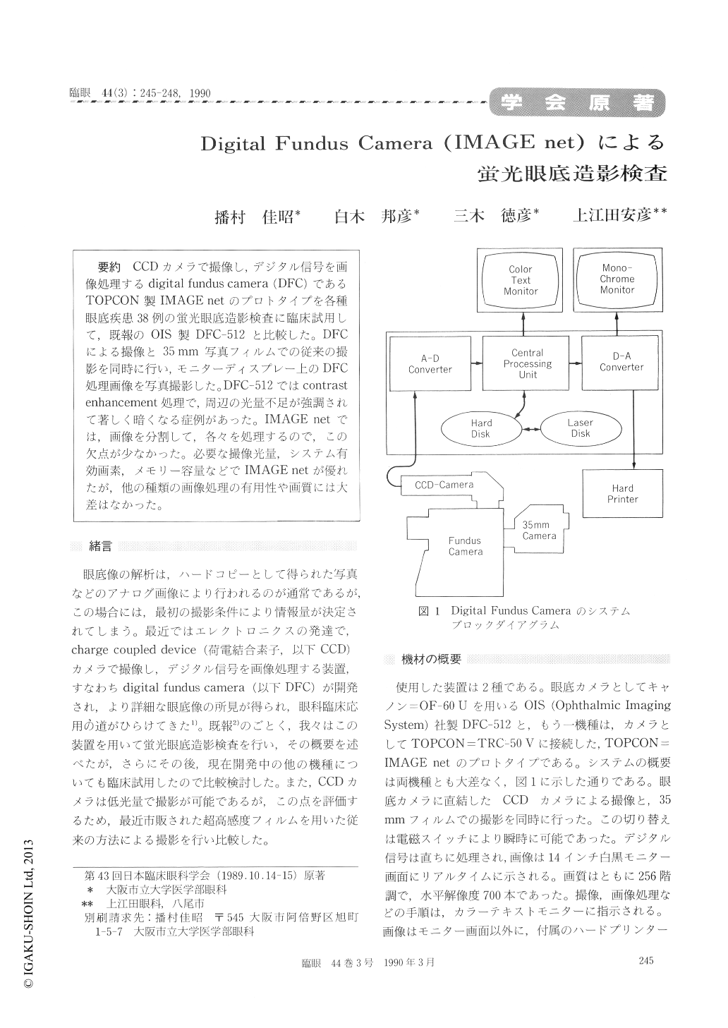
CCDカメラで撮像し,デジタル信号を画像処理するdigital fundus camera (DFC)であるTOPCON製IMAGE netのプロトタイプを各種眼底疾患38例の蛍光眼底造影検査に臨床試用して,既報のOIS製DFC−512と比較した。DFCによる撮像と35mm写真フィルムでの従来の撮影を同時に行い,モニターディスプレー上のDFC処理画像を写真撮影した。DFC−512ではcontrast enhancement処理で,周辺の光量不足が強調されて著しく暗くなる症例があった。IMAGE netでは,画像を分割して,各々を処理するので,この欠点が少なかった。必要な撮像光量,システム有効画素,メモリー容量などでIMAGE netが優れたが,他の種類の画像処理の有用性や画質には大差はなかった。
We evaluated the clinical feasibility of a new digital fundus camera, a prototype of TOPCON IMAGE net. The system utilizes a ; charge-coupled device camera, direct on-line acquisition, immedi-ate digitizing and manipulation. The quality of fluorescein angiograms with the system was infe-rior to those by conventional photographic means. Still, all the processed images were adequate for the diagnosis of fundus diseases and particularly in eyes with opaque media. Date accessibility and low -level flash power imaging were superior to theconventional methods.
The present system was as improved version of an earlier one, OIS DFC-512. Image processing with the earlier system produced an increase in contrast, often resulting in underexposure in the periphery of the images. The present system processed the image part by part. This segmental image process-ing masked any increase in contrast. It could proc-ess a variable intensity of enhancement by varying the number of segments. The whole image appear-ed as a mosaic pattern due to intensified contrast enhancements.

Copyright © 1990, Igaku-Shoin Ltd. All rights reserved.


