Japanese
English
- 有料閲覧
- Abstract 文献概要
- 1ページ目 Look Inside
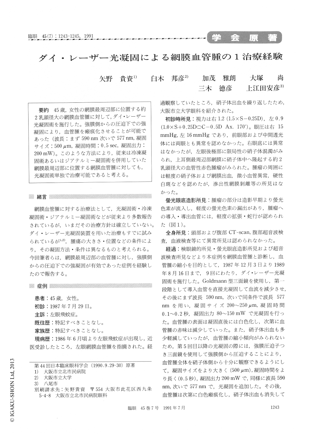
45歳,女性の網膜最周辺部に位置する約2乳頭径大の網膜血管腫に対して,ダイ・レーザー光凝固術を施行した。強膜側からの圧迫下での強凝固により,血管腫を瘢痕化させることが可能であった(波長:まず590nm次いで577nm,凝固サイズ:500μm,凝固時間:0.5sec,凝固出力:200mW)。このような方法により,従来は冷凍凝固術あるいはジアテルミー凝固術を併用していた網膜最周辺部に位置する網膜血管腫に対しても,光凝固術単独で治療可能であると考える。
A 45-year-female presented with myedesopsia inher left eye. Funduscopy revealed an oval vasculartumor, 2 disc diameters in size, in the far superiorperipheral retina. We treated the tumor with dyelaser photocoagulation under scleral indentation.We used wavelength of 590 nm followed by 577 nm,500 μm spot size, power output of 200mW andexposure time of 0.5sec. The present therapeuticmodality was effective in destroying the heman-gioma without recourse to cryoapplication or trans-scleral diathermy coagulation.

Copyright © 1991, Igaku-Shoin Ltd. All rights reserved.


