Japanese
English
- 有料閲覧
- Abstract 文献概要
- 1ページ目 Look Inside
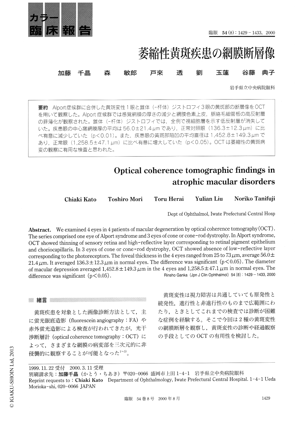
Alport症候群に合併した黄斑変性1眼と錐体(—杆体)ジストロフィ3眼の黄斑部の断層像をOCTを用いて観察した。Alport症候群では感覚網膜の厚さの減少と網膜色素上皮,脈絡毛細管板の高反射層の菲薄化が観察された。錐体(—杵体)ジストロフィでは,全例で視細胞層を示す低反射層が消失していた。疾患眼の中心窩網膜厚の平均は56.0±21.4μmであり,正常対照眼(136.3±12.3μm)に比べ有意に減少していた(p<0.01)。また,疾患眼の黄斑部陥凹の平均直径は1,452.8±149.3μmであり,正常眼(1,258.5±47.1μm)に比べ有意に増大していた(p<0.05)。OCTは萎縮性の黄斑病変の観察に有用な検査と思われた。
We examined 4 eyes in 4 patients of macular degeneration by optical coherence tomography (OCT). The series comprised one eye of Alport syndrome and 3 eyes of cone or cone-rod dystrophy. In Alport syndrome, OCT showed thinning of sensory retina and high-reflective layer corresponding to retinal pigment epithelium and choriocapillaris. In 3 eyes of cone or cone-rod dystrophy, OCT showed absence of low-reflective layer corresponding to the photoreceptors. The foveal thickness in the 4 eyes ranged from 25 to 73μm, average 56.0± 21.4μm. It averaged 136.3±12.3μm in normal eyes. The difference was significant (p<0.05). The diameter of macular depression averaged 1,452.8±149.3μm in the 4 eyes and 1,258.5±47.1μm in normal eyes. The difference was significant (p<0.05).

Copyright © 2000, Igaku-Shoin Ltd. All rights reserved.


