Japanese
English
- 有料閲覧
- Abstract 文献概要
- 1ページ目 Look Inside
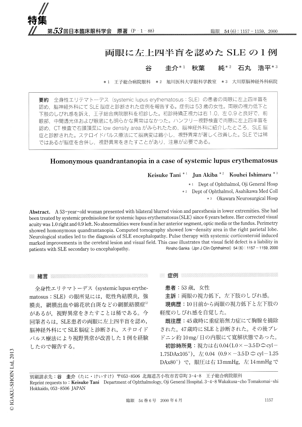
(P−1-88) 全身性エリテマトーデス(systemic lupus erythematosus:SLE)の患者の両眼に左上四半盲を認め,脳神経外科にてSLE脳症と診断された症例を報告する。症例は53歳の女性。両眼の視力低下と下肢のしびれ感を訴え,王子総合病院眼科を初診した。初診時矯正視力は右1.0,左0.9と良好で,前眼部,中間透光体および眼底にも明らかな異常はなかった。ハンフリー視野検査で両眼に左上四半盲を認め,CT検査で右頭頂葉にlow density areaがみられたため,脳神経外科に紹介したところ,SLE脳症と診断された。ステロイドパルス療法にて脳病変は縮小し,視野異常が著しく改善した。SLEでは稀ではあるが脳症を合併し,視野異常をきたすことがあり,注意が必要である。
A 53-year-old woman presented with bilateral blurred vision and paresthesia in lower extremities. She had been treated by systemic prednisolone for systemic lupus erythematosus (SLE) since 6 years before. Her corrected visual acuity was 1.0 right and 0.9 left. No abnormalities were found in her anterior segment, optic media or the fundus. Perimetry showed homonymous quandrantanopia. Computed tomography showed low-density area in the right parietal lobe. Neurological studies led to the diagnosis of SLE encephalopathy. Pulse therapy with systemic corticosteroid induced marked improvements in the cerebral lesion and visual field. This case illustrates that visual field defect is a liability in patients with SLE secondary to encephalopathy.

Copyright © 2000, Igaku-Shoin Ltd. All rights reserved.


