Japanese
English
- 有料閲覧
- Abstract 文献概要
- 1ページ目 Look Inside
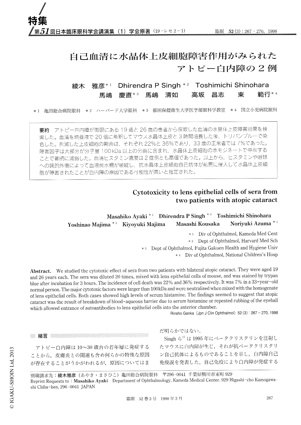
(19-レセ2-1) アトピー白内障が両眼にある19歳と26歳の患者から採取した血清の水晶体上皮障害効果を検索した。血清を培養液で20倍に希釈してマウス水晶体上皮と3時間培養した後,トリパンブルーで染色した。死滅した上皮細胞の割合は,それぞれ22%と36%であり,33歳の正常者では7%であった。障害因子は大部分が分子量100kDa以上の分画に含まれ,水晶体上皮細胞のホモジネートで中和することで著明に減弱した。血清ヒスタミン濃度は2症例とも高値であった。以上から,ヒスタミンや眼球への鈍的外傷によって血液房水柵が破綻し,抗水晶体上皮細胞自己抗体が前房に侵入して水晶体上皮細胞が障害されたことが白内障の原因である可能性が高いと推定された。
We studied the cytotoxic effect of sera from two patients with bilateral atopic cataract. They were aged 19 and 26 years each. The sera was diluted 20 times, mixed with lens epithelial cells of mouse, and was stained by trypan blue after incubation for 3 hours. The incidence of cell death was 22% and 36% respectively. It was 7% in a 33-year-old normal person. The major cytotoxic factors were larger than 100 kDa and were neutralized when mixed with the homogenate of lens epithelial cells. Both cases showed high levels of serum histamine. The findings seemed to suggest that atopic cataract was the result of breakdown of blood-aqueous barrier due to serum histamine or repeated rubbing of the eyeball which allowed entrance of autoantibodies to lens epithelial cells into the anterior chamber.

Copyright © 1998, Igaku-Shoin Ltd. All rights reserved.


