Japanese
English
特集 第49回日本臨床眼科学会講演集(3)
学会原著
Uveal effusionの強膜の組織学的検討
Histological findings of the sclera in uveal effusion
川端 紀穂
1
,
大谷 園子
1
,
村上 晶
1
,
水川 淳
1
,
沖坂 重邦
1
Kiho Kawabata
1
,
Sonoko Ooya
1
,
Akira Murakami
1
,
Atsushi Mizukawa
1
,
Shigekuni Okisaka
1
1防衛医科大学校眼科学教室
1Dept of Ophthalmol, National Defense Med Coll
pp.891-894
発行日 1996年5月15日
Published Date 1996/5/15
DOI https://doi.org/10.11477/mf.1410904889
- 有料閲覧
- Abstract 文献概要
- 1ページ目 Look Inside
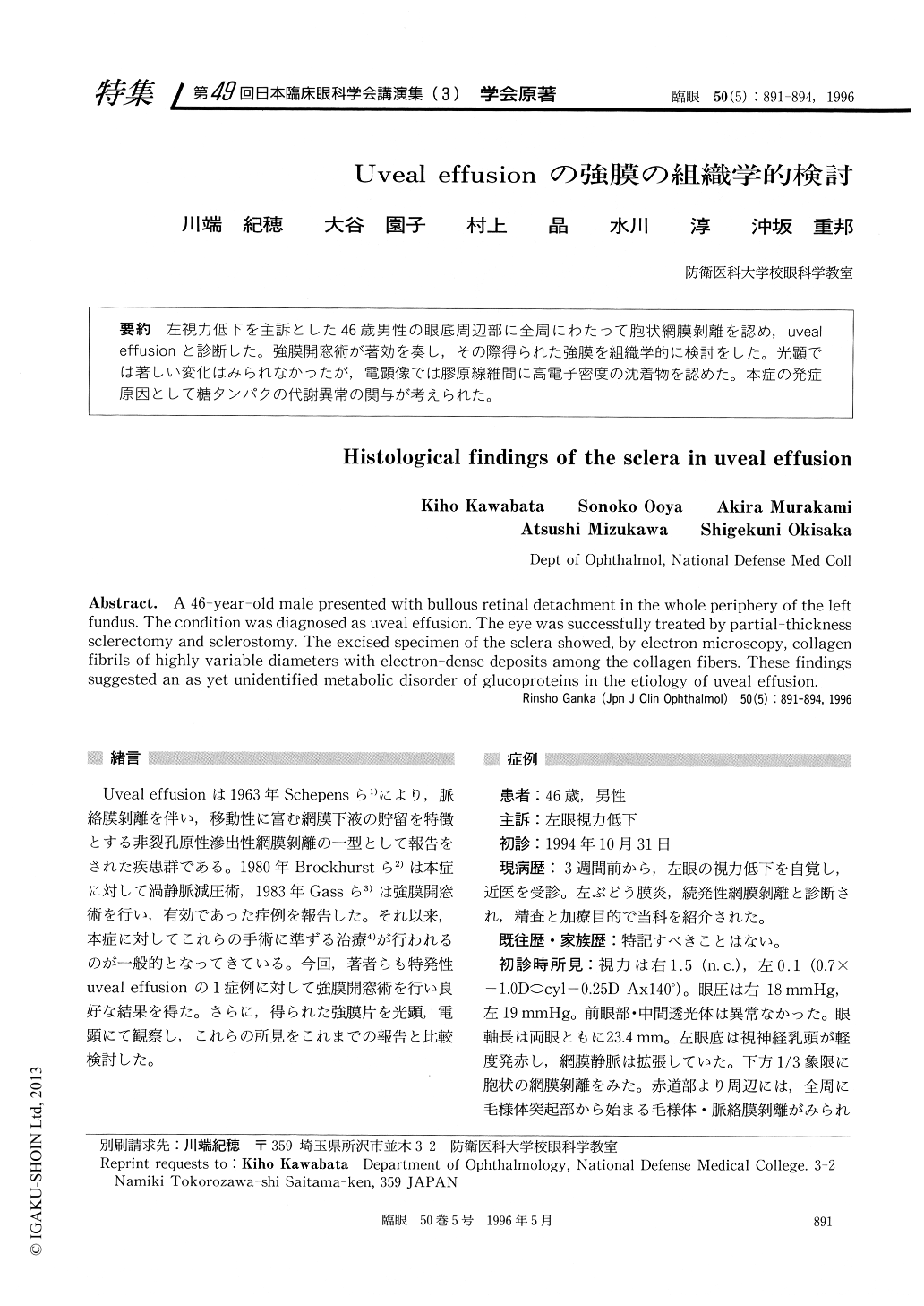
左視力低下を主訴とした46歳男性の眼底周辺部に全周にわたって胞状網膜剥離を認め,uvealeffusionと診断した。強膜開窓術が著効を奏し,その際得られた強膜を組織学的に検討をした。光顕では著しい変化はみられなかったが,電顕像では膠原線維間に高電子密度の沈着物を認めた。本症の発症原因として糖タンパクの代謝異常の関与が考えられた。
A 46-year-old male presented with bullous retinal detachment in the whole periphery of the left fundus. The condition was diagnosed as uveal effusion. The eye was successfully treated by partial-thickness sclerectomy and sclerostomy. The excised specimen of the sclera showed, by electron microscopy, collagen fibrils of highly variable diameters with electron-dense deposits among the collagen fibers. These findings suggested an as yet unidentified metabolic disorder of glucoproteins in the etiology of uveal effusion.

Copyright © 1996, Igaku-Shoin Ltd. All rights reserved.


