Japanese
English
- 有料閲覧
- Abstract 文献概要
- 1ページ目 Look Inside
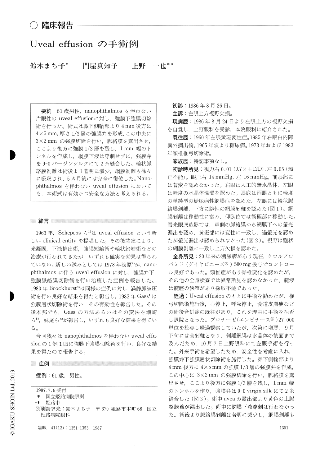
61歳男性,nanophthalmosを伴わない片眼性のuveal effusionに対し,強膜下強膜切除術を行った.術式は鼻下側輪部より4mm後方に4×5mm,厚さ1/3層の強膜弁を形成,この中央に3×2mmの強膜切除を行い,脈絡膜を露出させ,ここより後方に強膜1/3層を残し,1mm幅のトンネルを作成し,網膜下液は穿刺せずに,強膜弁を9-0バージンシルクにて2糸縫合した.輪状脈絡膜剥離は術後より著明に減少,網膜剥離も徐々に吸収され,5カ月後には完全に復位した.Nano-phthalmosを伴わないuveal effusionにおいても,本術式は有効かつ安全な方法と考えられる.
A 61-year-old male presented with annular ante-rior choroidal detachment associated with bullous retinal detachment in the inferior quadrant in his left eye. We diagnosed the condition as uveal effu-sion syndrome. The condition developed into total retinal detachment 6 weeks later after the initial refusal to surgery by the patient. We then perfor-med surgery to create a drainage channel from the choroid to the subconjunctival space.
A rectangular one-third thickness scleral trap-door, 4×5 mm in size, was made in the inferior nasal quadrant with its center 6 mm posterior to the limbus. A sclerostomy, 2 × 3 mm in size, was made in the center of the trapdoor. Posterior to the sclerostomy, a one-third thickness scleral tunnel was created. We made no attempt to perforate the choroid.
The retinal and choroidal detachment disappear-ed 5 months after surgery. The surgical procedure is advocated as a safe and effective one for uveal effusion.
Rinsho Ganka (Jpn J Clin Ophthalmol) 41(12) : 1351-1353,1987

Copyright © 1987, Igaku-Shoin Ltd. All rights reserved.


