Japanese
English
- 有料閲覧
- Abstract 文献概要
- 1ページ目 Look Inside
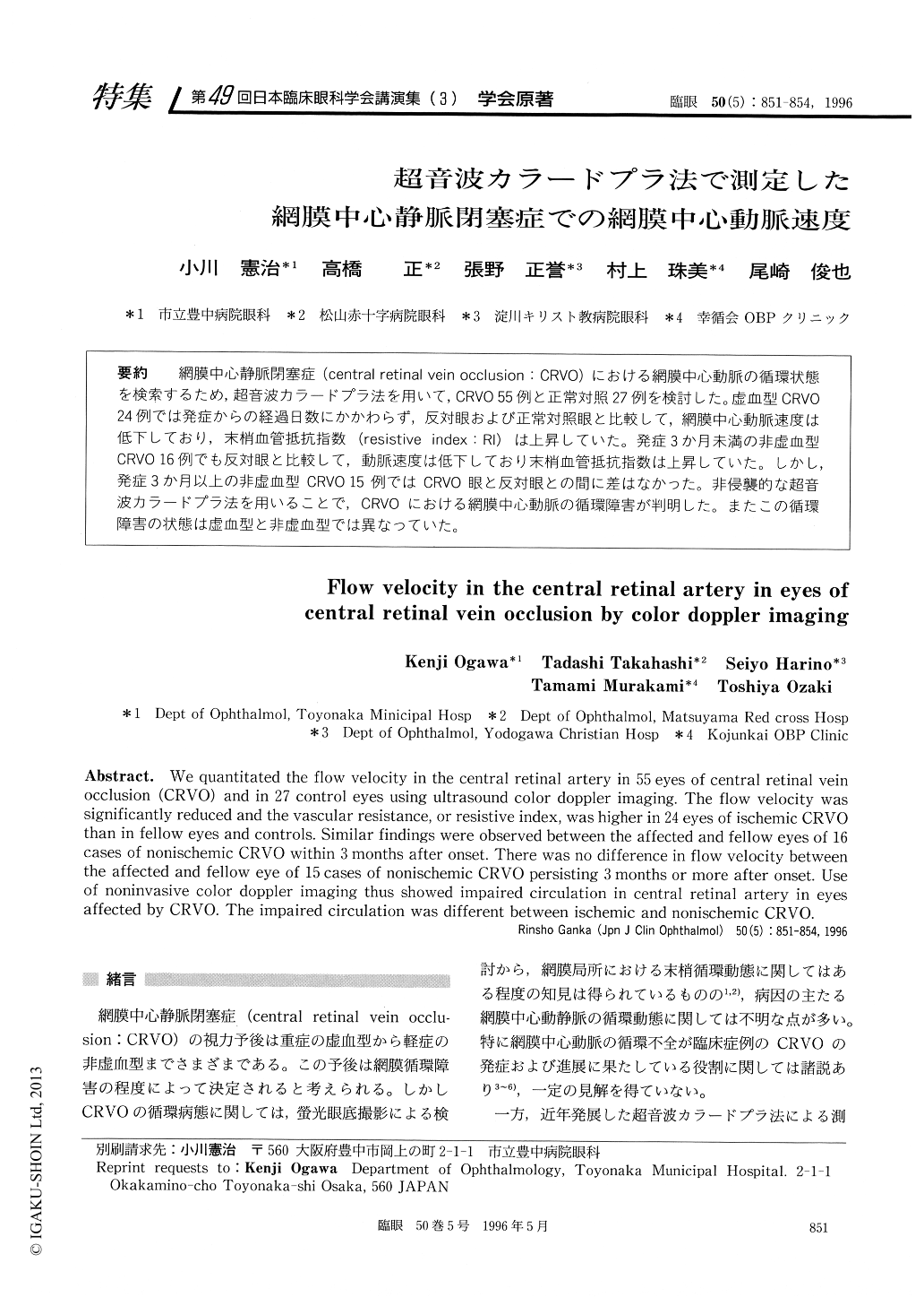
網膜中心静脈閉塞症(central retinal vein occsion:CRVO)における網膜中心動脈の循環状態を検索するため,超音波カラードプラ法を用いて,CRVO 55例と正常対照27例を検討した。虚血型CRVO24例では発症からの経過日数にかかわらず,反対眼および正常対照眼と比較して,網膜中心動脈速度は低下しており,末梢血管抵抗指数(resistive index:RI)は上昇していた。発症3か月未満の非虚血型CRVO 16例でも反対眼と比較して,動脈速度は低下しており末梢血管抵抗指数は上昇していた。しかし,発症3か月以上の非虚血型CRVO 15例ではCRVO眼と反対眼との間に差はなかった。非侵襲的な超音波カラードプラ法を用いることで,CRVOにおける網膜中心動脈の循環障害が判明した。またこの循環障害の状態は虚血型と非虚血型では異なっていた。
We quantitated the flow velocity in the central retinal artery in 55 eyes of central retinal vein occlusion (CRVO) and in 27 control eyes using ultrasound color doppler imaging. The flow velocity was significantly reduced and the vascular resistance, or resistive index, was higher in 24 eyes of ischemic CRVO than in fellow eyes and controls. Similar findings were observed between the affected and fellow eyes of 16 cases of nonischemic CRVO within 3 months after onset. There was no difference in flow velocity between the affected and fellow eye of 15 cases of nonischemic CRVO persisting 3 months or more after onset. Use of noninvasive color doppler imaging thus showed impaired circulation in central retinal artery in eyes affected by CRVO. The impaired circulation was different between ischemic and nonischemic CRVO.

Copyright © 1996, Igaku-Shoin Ltd. All rights reserved.


