Japanese
English
- 有料閲覧
- Abstract 文献概要
- 1ページ目 Look Inside
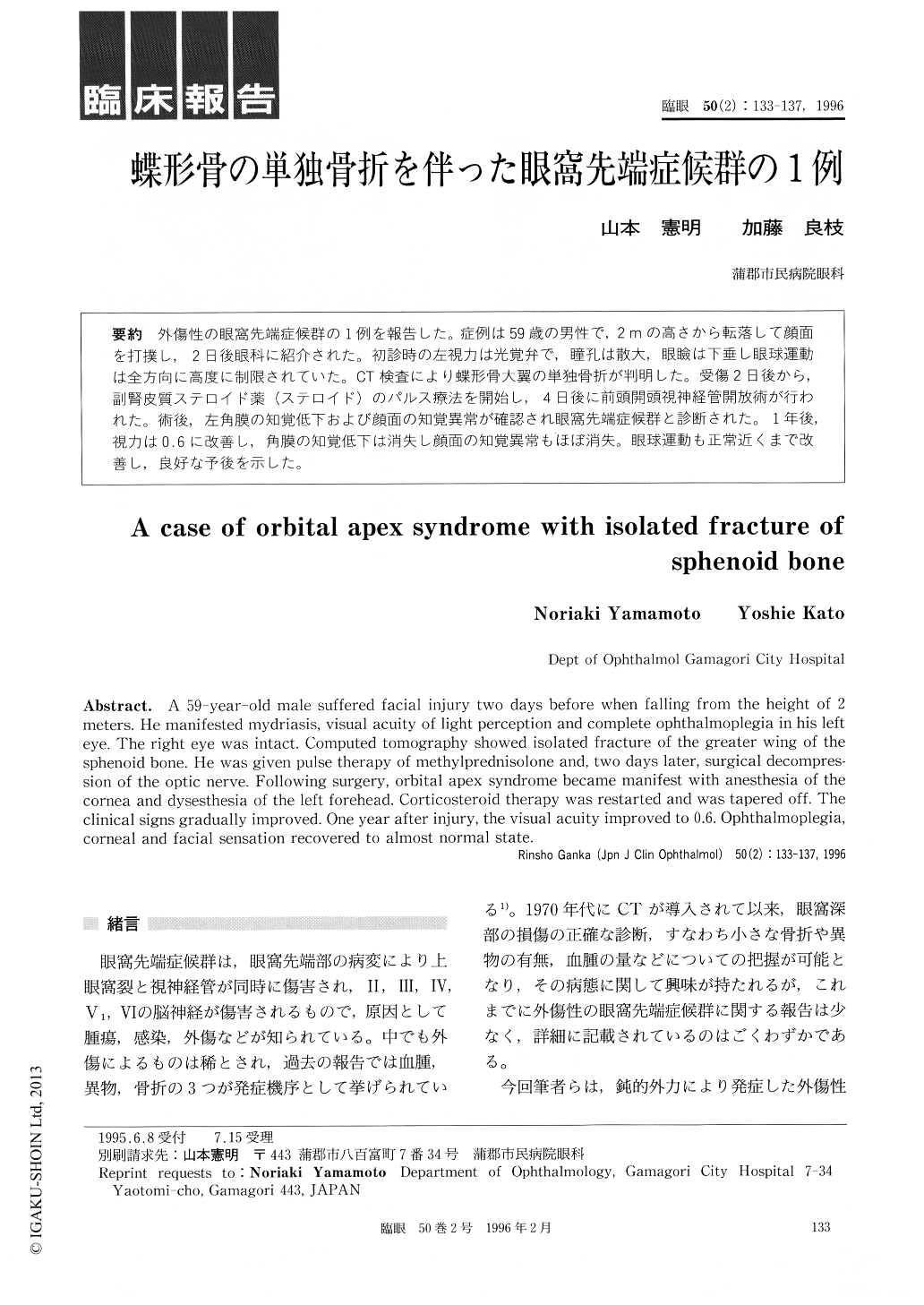
外傷性の眼窩先端症候群の1例を報告した。症例は59歳の男性で,2mの高さから転落して顔面を打撲し,2日後眼科に紹介された。初診時の左視力は光覚弁で,瞳孔は散大,眼瞼は下垂し眼球運動は全方向に高度に制限されていた。CT検査により蝶形骨大翼の単独骨折が判明した。受傷2日後から,副腎皮質ステロイド薬(ステロイド)のパルス療法を開始し,4日後に前頭開頭視神経管開放術が行われた。術後,左角膜の知覚低下および顔面の知覚異常が確認され眼窩先端症候群と診断された。1年後,視力は0.6に改善し,角膜の知覚低下は消失し顔面の知覚異常もほぼ消失。眼球運動も正常近くまで改善し,良好な予後を示した。
A 59-year-old male suffered facial injury two days before when falling from the height of 2 meters. He manifested mydriasis, visual acuity of light perception and complete ophthalmoplegia in his left eye. The right eye was intact. Computed tomography showed isolated fracture of the greater wing of the sphenoid bone. He was given pulse therapy of methylprednisolone and, two days later, surgical decompres-sion of the optic nerve. Following surgery, orbital apex syndrome became manifest with anesthesia of the cornea and dysesthesia of the left forehead. Corticosteroid therapy was restarted and was tapered off. The clinical signs gradually improved. One year after injury, the visual acuity improved to 0.6. Ophthalmoplegia, corneal and facial sensation recovered to almost normal state.

Copyright © 1996, Igaku-Shoin Ltd. All rights reserved.


