Japanese
English
- 有料閲覧
- Abstract 文献概要
- 1ページ目 Look Inside
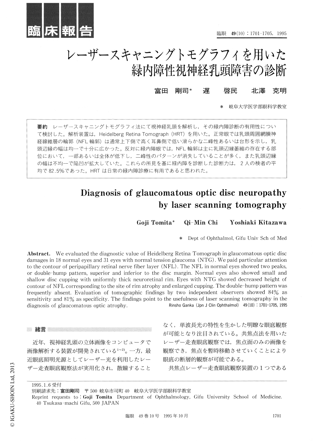
レーザースキャニングトモグラフィ法にて視神経乳頭を解析し,その緑内障診断の有用性について検討した。解析装置は,Heidelberg Retina Tomograph (HRT)を用いた。正常眼では乳頭周囲網膜神経線維層の輪郭(NFL輪郭)は通常上下側で高く耳鼻側で低い滑らかな二峰性あるいは台形を示し,乳頭辺縁の幅は均一で十分に広かった。反対に緑内障眼では,NFL輪郭は主に乳頭辺縁萎縮の存在する部位において,一部あるいは全体が低下し,二峰性のパターンが消失していることが多く,また乳頭辺縁の幅は不均一で陥凹が拡大していた。これらの所見を基に緑内障を診断した診断力は,2人の検者の平均で82.5%であった。HRTは日常の緑内障診療に有用であると思われた。
We evaluated the diagnostic value of Heidelberg Retina Tomograph in glaucomatous optic disc damages in 18 normal eyes and 31 eyes with normal tension glaucoma (NTG). We paid particular attention to the contour of peripapillary retinal nerve fiber layer (NFL). The NFL in normal eyes showed two peaks, or double-hump pattern, superior and inferior to the disc margin. Normal eyes also showed small and shallow disc cupping with uniformly thick neuroretinal rim. Eyes with NTG showed decreased height of contour of NFL corresponding to the site of rim atrophy and enlarged cupping. The double-hump pattern was frequently absent. Evaluation of tomographic findings by two independent observers showed 84% as sensitivity and 81% as specificity. The findings point to the usefulness of laser scanning tomography in the diagnosis of glaucomatous optic atrophy.

Copyright © 1995, Igaku-Shoin Ltd. All rights reserved.


