Japanese
English
- 有料閲覧
- Abstract 文献概要
- 1ページ目 Look Inside
- 参考文献 Reference
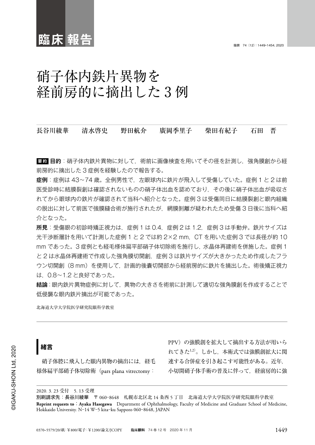
要約 目的:硝子体内鉄片異物に対して,術前に画像検査を用いてその径を計測し,強角膜創から経前房的に摘出した3症例を経験したので報告する。
症例:症例は43〜74歳。全例男性で,左眼球内に鉄片が飛入して受傷していた。症例1と2は前医受診時に結膜裂創は確認されないものの硝子体出血を認めており,その後に硝子体出血が吸収されてから眼球内の鉄片が確認されて当科へ紹介となった。症例3は受傷同日に結膜裂創と眼内組織の脱出に対して前医で強膜縫合術が施行されたが,網膜剝離が疑われたため受傷3日後に当科へ紹介となった。
所見:受傷眼の初診時矯正視力は,症例1は0.4,症例2は1.2,症例3は手動弁。鉄片サイズは光干渉断層計を用いて計測した症例1と2では約2×2mm,CTを用いた症例3では長径が約10mmであった。3症例とも経毛様体扁平部硝子体切除術を施行し,水晶体再建術を併施した。症例1と2は水晶体再建術で作成した強角膜切開創,症例3は鉄片サイズが大きかったため作成したフラウン切開創(8mm)を使用して,計画的後囊切開部から経前房的に鉄片を摘出した。術後矯正視力は,0.8〜1.2と良好であった。
結論:眼内鉄片異物症例に対して,異物の大きさを術前に計測して適切な強角膜創を作成することで低侵襲な眼内鉄片摘出が可能であった。
Abstract Purpose:To report three cases with intraocular iron foreign bodies which were safely removed from the anterior chamber through sclerocorneal tunnel after preoperative size measurement of the foreign bodies.
Case report:All cases were men aged from 43 to 74 years. Iron foreign bodies penetrated into the left eye in all the cases. In cases 1 and 2, while no conjunctival tears were found, fundus examination revealed vitreous hemorrhage at the former clinic. However, after spontaneous resolution of vitreous hemorrhage intraocular iron foreign bodies were detected and the patients were referred to our hospital. In case 3, scleral rupture and herniation of intraocular tissues had primarily been managed by scleral suturing at the former clinic on the day of injury, then he was referred to our hospital three days later due to possible retinal detachment.
Findings:Best corrected visual acuity of the injured eyes in the three cases ranged from 1.2 to hand motion. Sizes of the foreign bodies measured using optical coherence tomography were approximately 2×2 mm in cases 1 and 2, and approximately 10 mm in long diameter, estimated using computed tomography image, in case 3. In all three cases, combined pars plana vitrectomy and phacoemulsification and aspiration with intraocular lens implantation was performed. The foreign bodies were removed from the sclerocorneal tunnel for lens surgery in cases 1 and 2, whereas in case 3 the foreign body was removed from the frown incision(8 mm in size)due to its large size. Postoperative visual outcome was favorable, ranging from 0.8 to 1.2.
Conclusions:Preoperative size measurement and appropriate sclerocorneal tuunel construction were required to perform minimally-invasive procedures of intraocular iron foreign bodies.

Copyright © 2020, Igaku-Shoin Ltd. All rights reserved.


