Japanese
English
- 有料閲覧
- Abstract 文献概要
- 1ページ目 Look Inside
- 参考文献 Reference
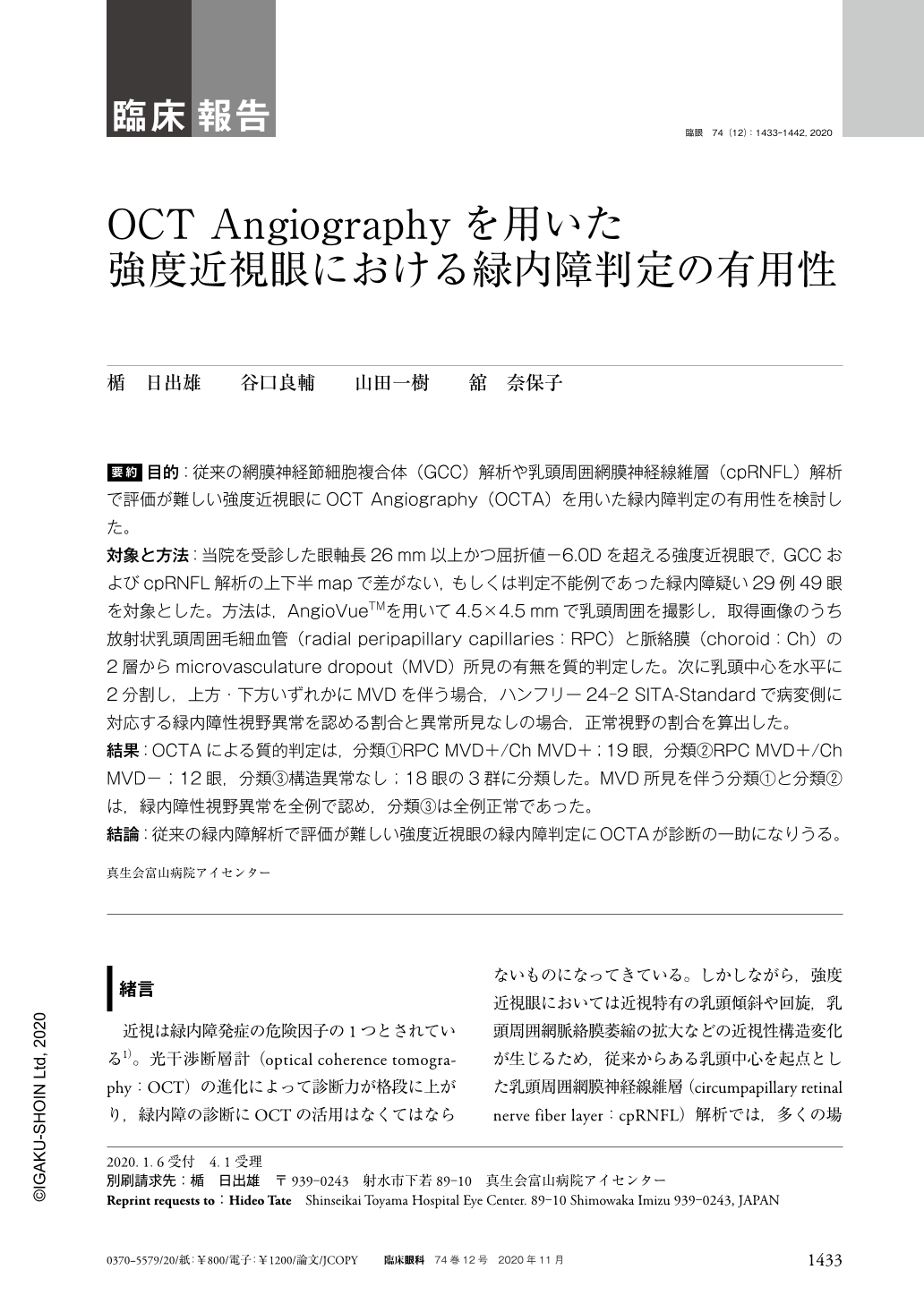
要約 目的:従来の網膜神経節細胞複合体(GCC)解析や乳頭周囲網膜神経線維層(cpRNFL)解析で評価が難しい強度近視眼にOCT Angiography(OCTA)を用いた緑内障判定の有用性を検討した。
対象と方法:当院を受診した眼軸長26mm以上かつ屈折値−6.0Dを超える強度近視眼で,GCCおよびcpRNFL解析の上下半mapで差がない,もしくは判定不能例であった緑内障疑い29例49眼を対象とした。方法は,AngioVueTMを用いて4.5×4.5mmで乳頭周囲を撮影し,取得画像のうち放射状乳頭周囲毛細血管(radial peripapillary capillaries:RPC)と脈絡膜(choroid:Ch)の2層からmicrovasculature dropout(MVD)所見の有無を質的判定した。次に乳頭中心を水平に2分割し,上方・下方いずれかにMVDを伴う場合,ハンフリー24-2 SITA-Standardで病変側に対応する緑内障性視野異常を認める割合と異常所見なしの場合,正常視野の割合を算出した。
結果:OCTAによる質的判定は,分類①RPC MVD+/Ch MVD+;19眼,分類②RPC MVD+/Ch MVD−;12眼,分類③構造異常なし;18眼の3群に分類した。MVD所見を伴う分類①と分類②は,緑内障性視野異常を全例で認め,分類③は全例正常であった。
結論:従来の緑内障解析で評価が難しい強度近視眼の緑内障判定にOCTAが診断の一助になりうる。
Abstract Purpose:To report the usefulness OCT Angiography(OCTA)of glaucoma diagnosis using in high myopia that an evaluation has difficult by conventional ganglion cell complex(GCC)or circumpapillary retinal nerve fiber layer(cpRNFL)analysis.
Subjects and methods:We evaluated 49 eyes of 29 glaucoma suspects visiting our hospital with an axial length of 26 mm or more and a refractive value greater than −6D. Using AngioVueTM to the case where there was no difference in the upper and lower half maps of the GCC and cpRNFL analysis, area where around the optic disc was photographed at 4.5×4.5 mm. The presence or absence of microvasculature dropout(MVD)findings was qualitatively judged from the two layers of radial peripapillary capillaries(RPC)and choroid(Ch)of the acquired images. If the center of the papilla is horizontally divided into two, accompanied by MVD either upward or downward, Humphrey 24-2 SITA-standard indicates that glaucomatous visual field abnormality corresponding to the lesion side is observed and abnormal findings are none percentage of visual field was calculated.
Results:The qualitative judgment by OCTA was classified into three groups of classification ① RPC MVD+/Ch MVD+ 19 eyes, classification ② RPC MVD+/Ch MVD− 12 eyes, classification ③ no abnormality 18 eyes. Classification ① and classification ② with abnormal findings showed glaucoma visual field abnormality in all cases, Classification ③ was all normal.
Conclusion:OCTA can help the diagnosis for a glaucoma decision of high myopia that an evaluation has difficult by conventional glaucoma analysis.

Copyright © 2020, Igaku-Shoin Ltd. All rights reserved.


