Japanese
English
- 有料閲覧
- Abstract 文献概要
- 1ページ目 Look Inside
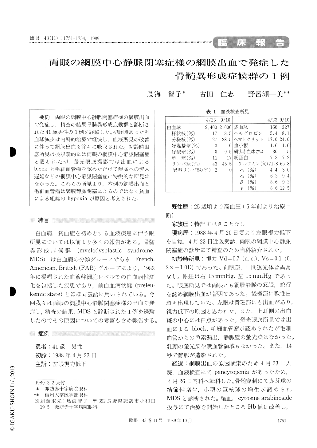
両眼の網膜中心静脈閉塞症様の網膜出血で発症し,精査の結果骨髄異形成症候群と診断された41歳男性の1例を経験した。初診時あった汎血球減少は内科的治療で軽快し,血液所見の改善に伴って網膜出血も徐々に吸収された。初診時眼底所見は検眼鏡的には両眼の網膜中心静脈閉塞症と思われたが,螢光眼底撮影では出血によるblockと毛細血管瘤を認めただけで静脈への流入遅延などの網膜中心静脈閉塞症に特徴的な所見はなかった。これらの所見より,本例の網膜出血と毛細血管瘤は網膜静脈閉塞によるのではなく貧血による組織のhypoxiaが原因と考えられた。
A 41-year-old male presented with blurring of vision in his left eye since 2 days before. He had been suffering from systemic hypertension since the age of 25. Funduscopy revealed bilateral retinal hemorrhage associated with dilated retinal veins and soft exudates. The fundus features closely simulated those of central retinal vein occlusion. Fluorescein showed the presence of numerous capil-lary microaneurysms but showed no leakage of dyefrom retinal vessels, dye staining of vessel wall, or fillig delay. Hematological studies showed pan-cytopenia leading to the diagnosis of myelodysplas-tic syndrome. Systemic treatments with blood transfusion and cytosine arabinoside preparations resulted in improvements in hematological findings and fundus findings. Full recovery of visual acuity was attained 5 months later. The observed retinal hemorrhage and capillary microaneurysms seemed to be due to anemia and not to retinal vein occlu-sion.
Copyright © 1989, Igaku-Shoin Ltd. All rights reserved.