Japanese
English
- 有料閲覧
- Abstract 文献概要
- 1ページ目 Look Inside
41歳男性の右眼窩内に発生した動脈瘤様骨嚢腫(aneurysmal bone cyst)の1例を報告した。
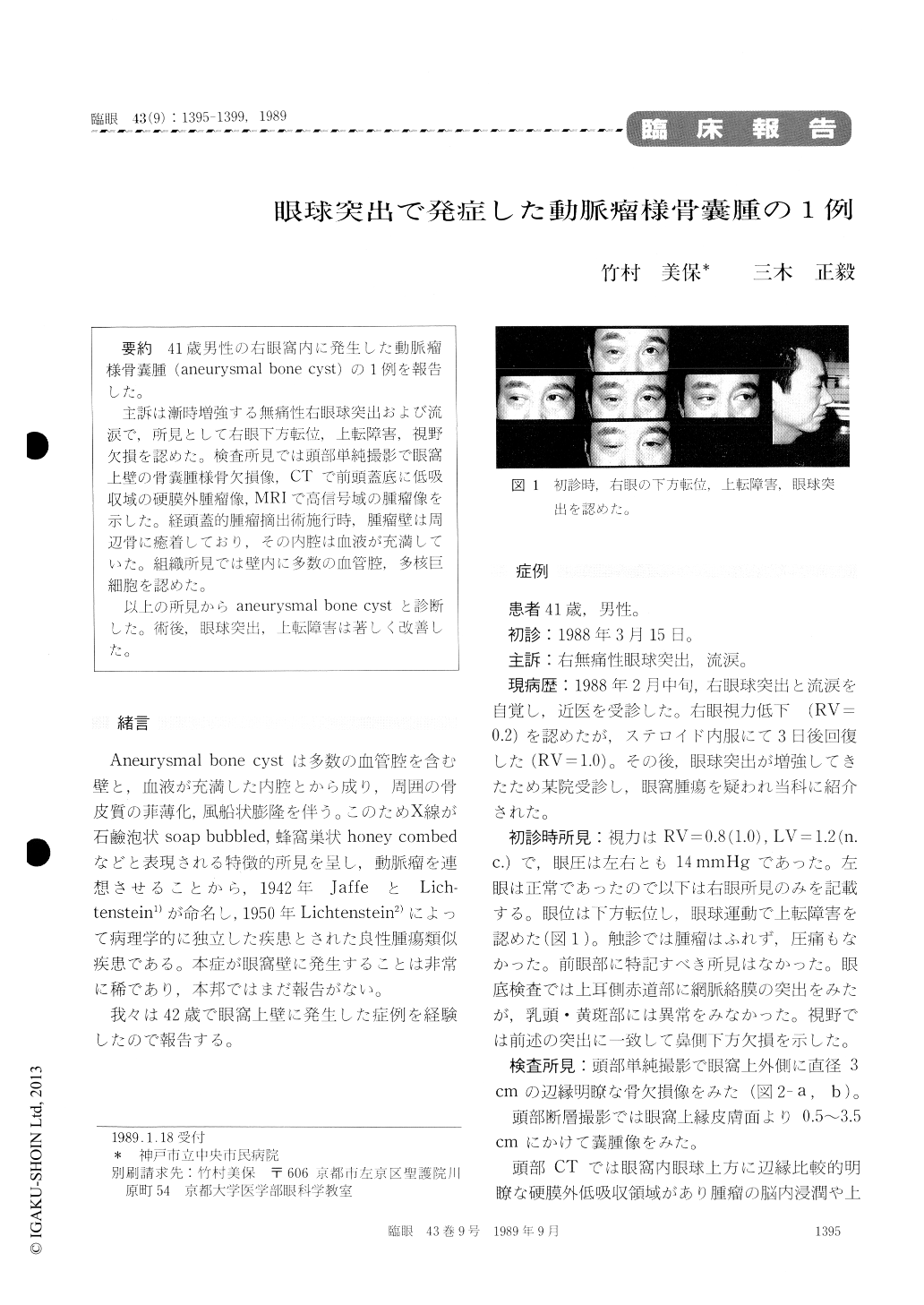
主訴は漸時増強する無痛性右眼球突出および流涙で,所見として右眼下方転位,上転障害,視野欠損を認めた。検査所見では頭部単純撮影で眼窩上壁の骨嚢腫様骨欠損像,CTで前頭蓋底に低吸収域の硬膜外腫瘤像,MRIで高信号域の腫瘤像を示した。経頭蓋的腫瘤摘出術施行時,腫瘤壁は周辺骨に癒着しており,その内腔は血液が充満していた。組織所見では壁内に多数の血管腔,多核巨細胞を認めた。
以上の所見からaneurysmal bone cystと診断した。術後,眼球突出,上転障害は著しく改善した。
A 41-year-old man presented with progressive proptosis and downward displacement of the right eye of one month's duration. Roentgenogram and computed tomography showed an extradural mass in the roof of the right orbit with bone erosion. Magnetic resonance imaging (MRI), showed by T1, and T2 imaging, showed the mass to be of high signal intensity. Transcranial extirpation of the encapsulated tumor led to the diagnosis of aneurys-mal bone cyst of the orbit. Histopathologically, the capsule multinucleated giant cells and non-endoth-elial vascular channels. Surgery was followed by improvements in eye position and movement of extraocular muscles. To the best of our knowledge, this is the first reported case of aneurysmal bone cyst of the orbit in Japan. MRI findisgs were crucial in the etiological diagnosis of proptosis.

Copyright © 1989, Igaku-Shoin Ltd. All rights reserved.


