Japanese
English
- 有料閲覧
- Abstract 文献概要
- 1ページ目 Look Inside
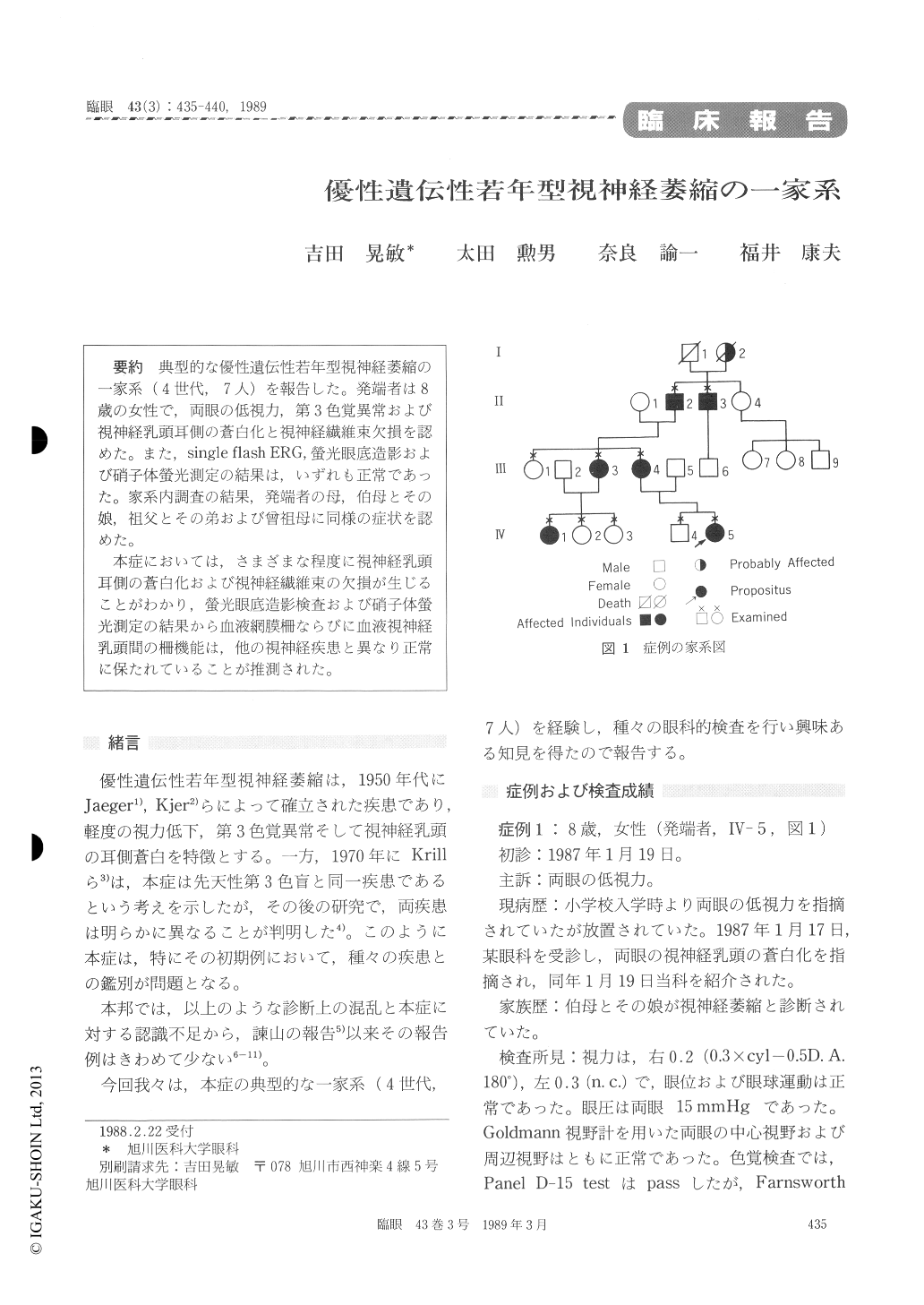
典型的な優性遺伝性若年型視神経萎縮の一家系(4世代,7人)を報告した。発端者は8歳の女性で,両眼の低視力,第3色覚異常および視神経乳頭耳側の蒼白化と視神経繊維束欠損を認めた。また,single flash ERG,螢光眼底造影および硝子体螢光測定の結果は,いずれも正常であった。家系内調査の結果,発端者の母,伯母とその娘,祖父とその弟および曾祖母に同様の症状を認めた。
本症においては,さまざまな程度に視神経乳頭耳側の蒼白化および視神経繊維束の欠損が生じることがわかり,螢光眼底造影検査および硝子体螢光測定の結果から血液網膜柵ならびに血液視神経乳頭間の柵機能は,他の視神経疾患と異なり正常に保たれていることが推測された。
We observed a pedigree with typical type of dominant juvenile optic atrophy consisting of 7 patients over 4 generations. A female child of 8 years, the propositus, presented with impaired visual acuity in both eyes. She manifested temporal pallor of the optic disc and difficulty in color discrimination compatible with tritanomaly. Find-ings with single flash ERG, fluorencein angiogra-phy and vitreous fluorophotometry were within normal range. Essentially similar findings were detected through examination of members in the pedigree : her mother, aunt and her daughter,grandfather and his brother, and great-grand-mother on the paternal side.
All the affected members in the pedigree manifested varying degrees of either temporal or total optic disc pallor. All the members showed normal peripheral visual field. The visual acuity ranged from 0.1 to 0.4. Findings with fluorescein fundus angiography and vitreous fluorophotometry indicated that the blood-retinal and blood-disc barriers were intact: a unique feature when compared to other optic neuropathies.

Copyright © 1989, Igaku-Shoin Ltd. All rights reserved.


