Japanese
English
- 有料閲覧
- Abstract 文献概要
- 1ページ目 Look Inside
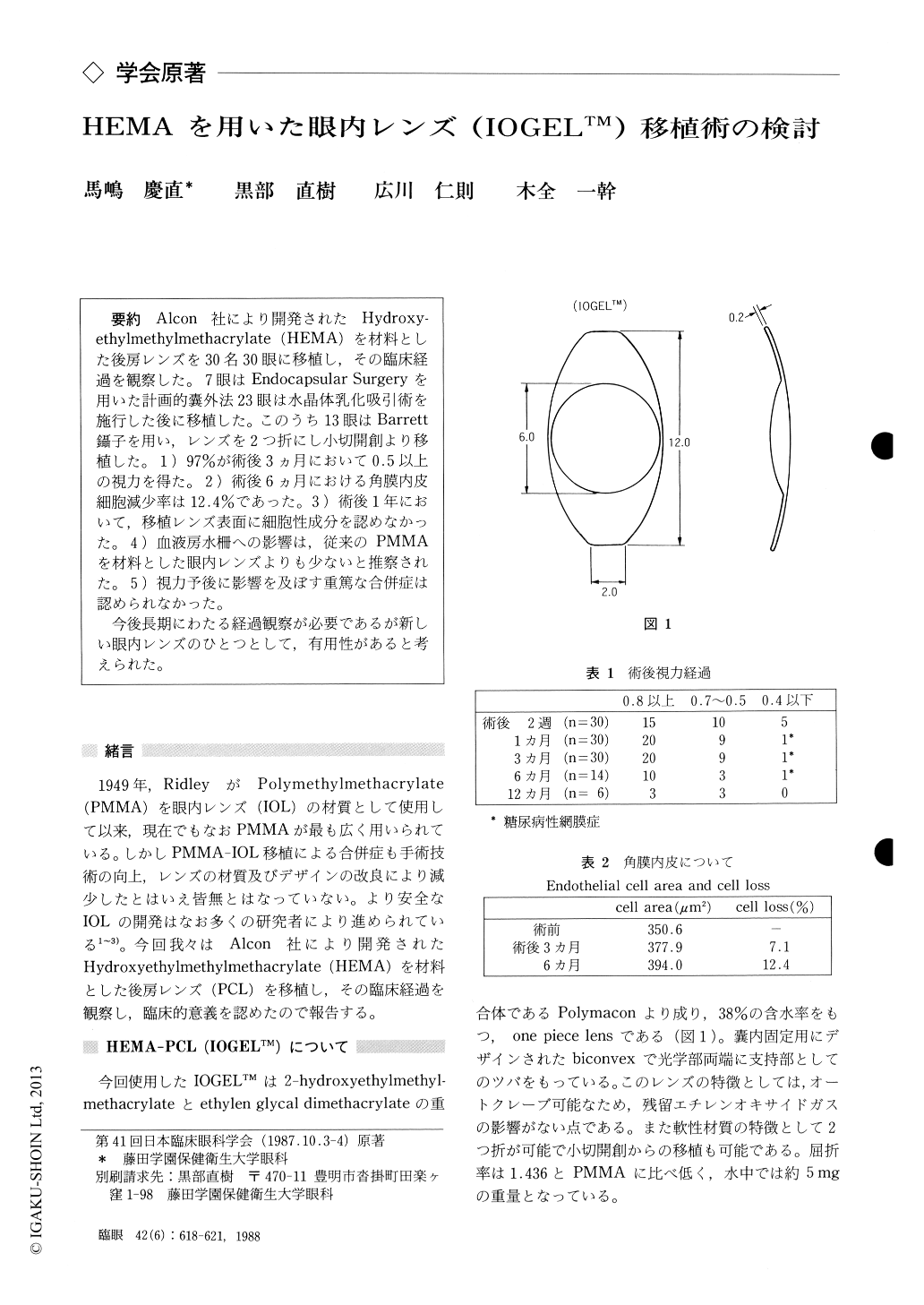
Alcon社により開発されたHydroxy-ethylmethylmethacrylate (HEMA)を材料とした後房レンズを30名30眼に移植し,その臨床経過を観察した.7眼はEndocapsular Surgeryを用いた計画的嚢外法23眼は水晶体乳化吸引術を施行した後に移植した.このうち13眼はBarrett鑷子を用い,レンズを2つ折にし小切開創より移植した.1)97%が術後3カ月において0.5以上の視力を得た.2)術後6カ月における角膜内皮細胞減少率は12.4%であった.3)術後1年において,移植レンズ表面に細胞性成分を認めなかった.4)血液房水柵への影響は,従来のPMMAを材料とした眼内レンズよりも少ないと推察された.5)視力予後に影響を及ぼす重篤な合併症は認められなかった.
今後長期にわたる経過観察が必要であるが新しい眼内レンズのひとつとして,有用性があると考えられた.
We performed implantation surgery in 30 eyes with posterior chamber intraocular lens (IOL) made of hydroxyethylmethylmethacrylate (HEMA), commercially available in U. S. A. as IOGELTM. The IOL contents water up to 38% of its weight, is pliable and withstand autoclaving. We performed the IOL implantation following extracapsular cataract extraction (ECCE) using endocapsular technique in 7 eyes, and following phacoemulsification/aspiration (KPE) in 23. In the KPE series, the lens was inserted held with Gaskin forceps through a 5 mm opening in 13 eyes, and was folded with Barrett forceps through 4.5 to 5 mmopening in 10 eyes.
At an evaluation 3 months after surgery, the visual acuity of 20/40 or better was attained in all the eyes but one (97%). Specular microscopic studies showed an endothelial cell loss of 7.1% at 3 months after surgery and of 12.4% at 6 months. When examined by specular microscopy one year after surgery, the surface of the IOL remained clear and was free of adherent cell-like structures. Fluor-ophotometric studies showed lesser damage to the blood-aqueous barrier when the IOL was implanted in the bag than out of the bag. As an overall estimation, the amount of damage to the blood -aqueous barrier in the present series was the least throughout a larger series of pseudophakic eyes evaluated by us. No major complications were observed in the present study.
Rinsho Ganka (Jpn J Clin Ophthalmol) 42(6) : 618-621, 1988

Copyright © 1988, Igaku-Shoin Ltd. All rights reserved.


