Japanese
English
- 有料閲覧
- Abstract 文献概要
- 1ページ目 Look Inside
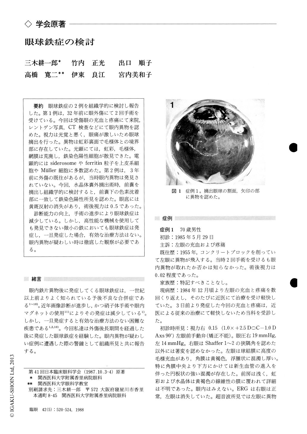
眼球鉄症の2例を組織学的に検討し報告した.第1例は,32年前に眼外傷にて2回手術を受けている.今回は受傷眼の充血と疼痛にて来院,レントゲン写真,CT検査などにて眼内異物を認めた.視力は光覚と悪く,眼痛が激しいため眼球摘出を行った.異物は虹彩裏面で毛様体との境界部に存在していた.光顕にては,虹彩,毛様体,網膜は荒廃し,鉄染色陽性細胞が散見できた.電顕的にはsiderosomeやferritin粒子を上皮系細胞やMüller細胞に多数認めた.第2例は,3年前に外傷の既往があるが,当時眼内異物は発見されていない.今回,水晶体嚢外摘出術時,前嚢を摘出し組織学的に検討すると,前嚢下の色素沈着部に一致して鉄染色陽性所見を認めた.眼底には黄斑反射の消失があり,術後視力は0.5であった.
診断能力の向上,手術の進歩により眼球鉄症は減少している.しかし,高性能な機械を使用しても発見できない微小の鉄においても眼球鉄症は発症し,一旦発症した場合,有効な治療方法はない.眼内異物が疑わしい時は徹底した観察が必要である.
We observed 2 cases of ocular siderosis. The first case, a 70-year-old man, developed severe pain in his left eye 32 years after perforating injury. X-ray studies showed a foreign body in the anterior cham-ber behind the opaque cornea. The eye was enu-cleated because of intractable pain. Histopath-ologically, the ciliary body and retina showed marked atrophy. Siderosomes and ferritin particles were dispersed throughout the eyeball, particularlyin the epithelial cells of the iris and the ciliary body, retinal pigment epithelium, and Mueller cells.
The second case, a 52-year-old woman, devel-oped cataract after ocular injury 3 years before. The cornea and iris showed signs of penetrating injury. Rust spots were seen on the anterior surface of cataractous lens. We obtained the anterior cap-sule during surgery for cataract. Histopathological-ly, we observed proliferation of subcapsular lens epithelium underlying the rust spots with positive iron reaction by Berlin blue staining.
Rinsho Ganka (Jpn J Clin Ophthalmol) 42(5) : 520-524, 1988

Copyright © 1988, Igaku-Shoin Ltd. All rights reserved.


