Japanese
English
- 有料閲覧
- Abstract 文献概要
- 1ページ目 Look Inside
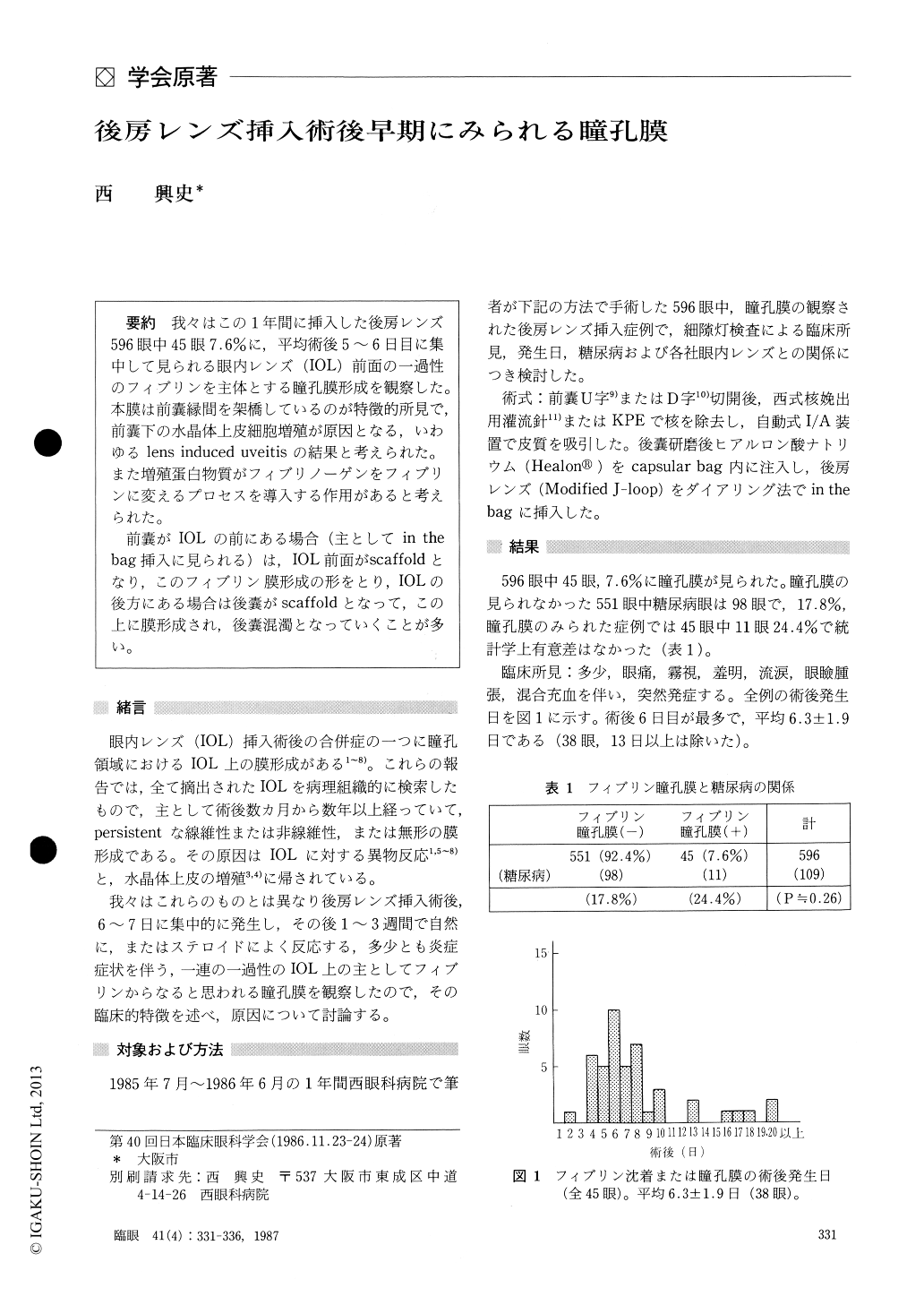
我々はこの1年間に挿入した後房レンズ596眼中45眼7.6%に,平均術後5〜6日目に集中して見られる眼内レンズ(IOL)前面の一過性のフィブリンを主体とする瞳孔膜形成を観察した.本膜は前嚢縁間を架橋しているのが特徴的所見で,前嚢下の水晶体上皮細胞増殖が原因となる,いわゆるlens induced uveitisの結果と考えられた.また増殖蛋白物質がフィブリノーゲンをフィブリンに変えるプロセスを導入する作用があると考えられた.
前嚢がIOLの前にある場合(主としてin thebag挿入に見られる)は,IOL前面がscaffoldとなり,このフィブリン膜形成の形をとり,IOLの後方にある場合は後嚢がscaffoldとなって,この上に膜形成され,後嚢混濁となっていくことが多い.
We observed transient formation of pupillary membrane in 45 out of a series of 596 eyes (7.6%) who received posterior chamber lens implantation during the foregoing one-year period. The mem-brane was adherent to the anterior surface of theintraocular lens (IOL) and seemed to consist of fibrin. The condition usually occurred 5 to 6 days after surgery. This membrane characteristically filled the space between the IOL surface and the anterior capsule, suggesting a lens-induced uveitis due to proliferation of epithelial cells of the lens under the anterior capsule. Probably, the proteins produced during cell proliferation initiated the con-version of fibrinogen into fibrin.
When the anterior capsule is positioned anteriorto the IOL, a situation frequently observed after in-the-bag insertion, the anterior surface of the IOL acts as a scaffold, facilitating the formation of this fibrin membrane. When the anterior capsule is positioned posterior to the IOL, a similar membrane may be formed on the posterior capsule, which actsas a scaffold, resulting in posterior capsule ーpacifi-cation.
Rinsho Ganka (Jpn J Clin Ophthalmol) : 41(4) : 331-336, 1987

Copyright © 1987, Igaku-Shoin Ltd. All rights reserved.


