Japanese
English
- 有料閲覧
- Abstract 文献概要
- 1ページ目 Look Inside
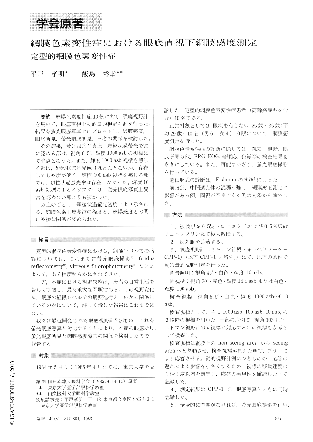
網膜色素変性症10例に対し,眼底視野計を用いて,眼底直視下動的量的視野計測を行った.結果を螢光眼底写真上にプロットし,網膜感度,眼底所見,螢光眼底所見,三者の関係を検討した.
その結果,螢光眼底写真上,顆粒状過螢光を密に認める部は,視角6,5',輝度1000asbの視標にて暗点となった.また,輝度1000asb視標を感じる部は,顆粒状過螢光像はほとんどないか,存在しても密度が低く,輝度100asb視標を感じる部では,顆粒状過螢光像は存在しなかった.輝度10asb視標によるイソプターは,螢光眼底写真上異常を認めない部よりも狭かった.
以上のごとく,顆粒状過螢光密度により示される,網膜色素上皮萎縮の程度と,網膜感度との間に密接な関係が認められた.
We performed campimetry in 10 cases with typical primary pigmentary retinal dystrophy under simultane-ous observation of the fundus with the use of fundus photoperimeter. The campimetric records were mat-ched against fluorescein angiogram by superimposing to evaluate the correlation between retinal sensitivity and fluorescein findings.
The retinal sensitivity was closely associated with thedensity of granular hyperfluorescence in the fluorescein angiogram resulting from atrophy of the retinal pig-ment epithelium. High-density granular hyperfluore-scent areas failed to recognize the bright test target (1000 asb). Retinal areas sensitive to 100 asb target lacked granular hyperfluorescence. Those sensitive to 1000 asb alone manifested low-density granular hyper-fluorescence. The isopter for 10 asb target was gene-rally smaller than retinal areas with no abnormalities on fluorescein angiogram.
Rinsho Ganka (Jpn J Clin Ophthalmol) 40(8) : 877-881, 1986

Copyright © 1986, Igaku-Shoin Ltd. All rights reserved.


