Japanese
English
- 有料閲覧
- Abstract 文献概要
- 1ページ目 Look Inside
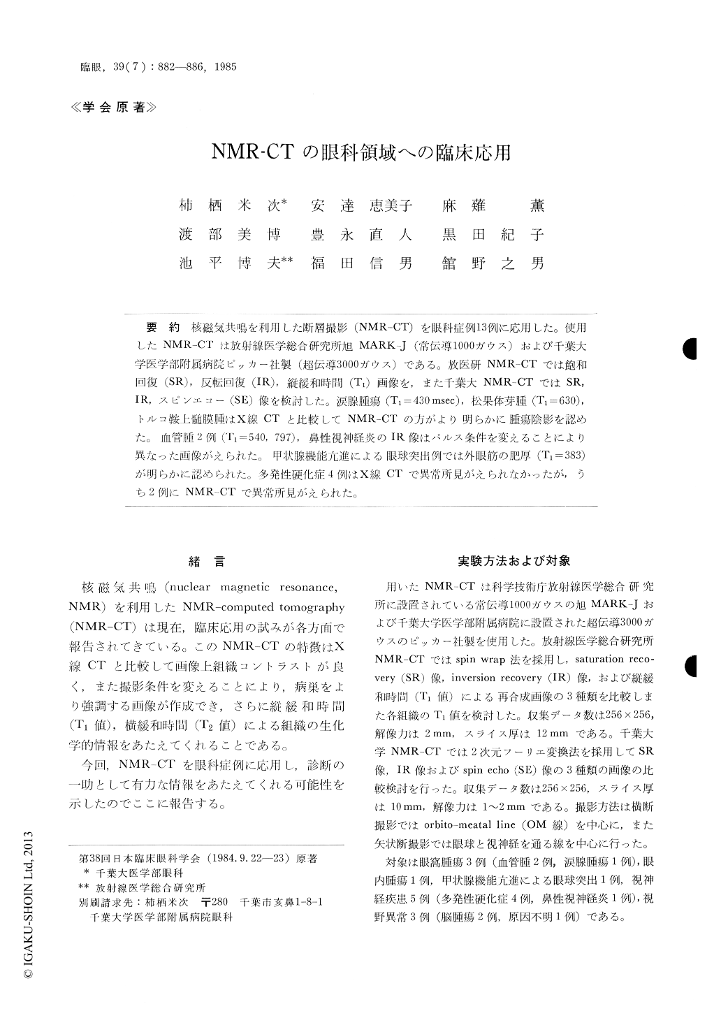
核磁気共鳴を利用した断層撮影(NMR-CT)を眼科症例13例に応用した.使用したNMR-CTは放射線医学総合研究所旭MARK-J (常伝導1000ガウス)および千葉大学医学部附属病院ピッカー社製(超伝導3000ガウス)である.放医研NMR-CTでは飽和回復(SR),反転回腹(IR),縦緩和時間(T1)画像を,また千葉大NMR-CTではSR,IR,スピンエコー(SE)像を検討した.涙腺腫瘍(T1=430msec),松果体芽腫(T1=630),トルコ鞍上髄膜腫はX線CTと比較してNMR-CTの方がより明らかに腫瘍陰影を認めた.血管腫2例(T1=540,797),鼻性視神経炎のIR像はパルス条件を変えることにより異なった画像がえられた.甲状腺機能亢進による眼球突出例では外限筋の肥厚(T1=383)が明らかに認められた.多発性硬化症4例はX線CTで異常所見がえられなかったが,うち2例にNMR-CTで異常所見がえられた.
We examined 13 patients with eye-related diseases with the use of magnetic resonance computed tomo-graphy (NMR-CT). The findings were evaluated by saturation recovery (SR), inversion recovery (IR), spin echo (SE) and T1 as parameters. The cases included 3 cases of orbital tumor, 1 intraocular tumor, 1 dysthyroid exophthalmos, 5 cases of optic nerve affections and 3 cases with impaired visual field.
When compared with conventional computed axi-al tomography (CT), NMR imaging showed clear-arer images of the tumor in pineoblastoma, supra-sellar meningioma and lacrimal gland tumor. The swelling of extraocular muscles in dysthyroid ex-ophthalmos could be demonstrated by T1 imaging. We studied the significance of Ti value in various tissues and under normal and pathological condi-tions. We reached the conclusion that the T1 value is a fair indicator for the pathological natures of tumors.

Copyright © 1985, Igaku-Shoin Ltd. All rights reserved.


