Japanese
English
- 有料閲覧
- Abstract 文献概要
- 1ページ目 Look Inside
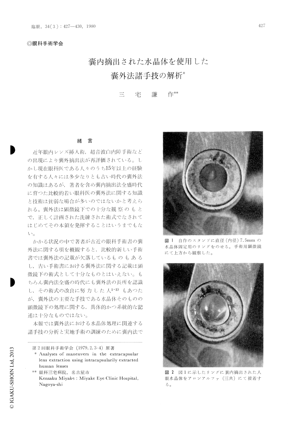
嚢内摘出された水晶体を使用して,嚢外法における前嚢切除,核摘出および後嚢研麿の3手技を行つてみた。当法では可視性が生体眼で行うよりすぐれており,各手技の実態が把握でき,生体眼における実際の手技の訓練と分析に役立つ。
前嚢切開は一気に行わず小刻みな小切開孔をミシン目状に作製し,後に全体に切開線を広げる方法が最も安全であつた。核の摘出は予めチストトームかdisposable needleで核を刺入し,水晶体の中央を中心にかるい円運動をするか上下左右にかるく運動させることにより皮質部における後嚢と核の接着を離断することにより容易になされる。また後嚢はScratcherによる研麿にたえる強靱な膜であることが分つた。さらに後嚢研麿により残留皮質はある程度除去しうることが直視下で観察できた。
Using intracapsularily extracted human lenses, anterior capsulectomy, removal of the lens nucleus and polishing of the posterior lens capsule were performed. As "in vitro" model of extracapsular lens extraction, this method was found to have much better visibility than actual "in vivo" ope-ration, and was useful to analyze as well to practice the key points of above techniques. Some details obtained from this method are as follows.
Anterior capsulotomy should not be done in one session, but should be designed as dottled line a tfirst, followed by continued line after-wards. The nucleus is removed by buried disposable needle or tip of the cystotome. Several types of junctions between the nucleus and the posterior capsule are solved by slow circular, vertical or horizontal actions of the buried needle or tip of the cystotome. 'rite posterior capsule was found strong enough to tolerate the mechanical stimulus by the scratching needle. Further, it was recog-nized under high magnification that polishing was effective to remove the residual lens cell processes to some extent.

Copyright © 1980, Igaku-Shoin Ltd. All rights reserved.


