Japanese
English
- 有料閲覧
- Abstract 文献概要
- 1ページ目 Look Inside
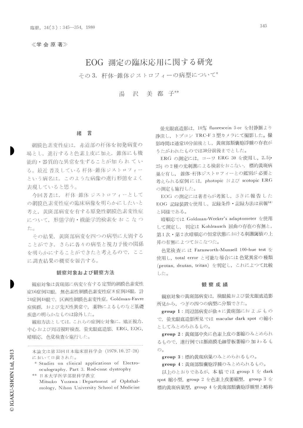
黄斑部病変を有する原発性網膜色素変性症24症例48眼を,杵体−錐体ジストロフィーという疾病概念にたつて,検眼鏡および螢光眼底造影所見,眼球電図(EOG),網膜電図(ERG),色覚検査などの所見によつて,下記の四つの病型に分類できるものと考える。
(1)黄斑部dark spot縮小型:周辺部病変が徐々に黄斑部におよぶもので,螢光眼底造影ではmacular dark spotの縮小としてみとめられるもの。
(2)黄斑部色素上皮萎縮型:黄斑部中央に色素上皮の萎縮のみとめられるもので,進行例では脈絡膜毛細管板萎縮の加わるもの。
(3)標的黄斑病巣型
(2)および(3)はabiotrophicな変化が黄斑部にもあつて出現する病型と考えられる。
(4)黄斑部類嚢胞浮腫型:黄斑部類嚢胞浮腫のみとめられるもの。ジストロフィー過程で生ずる血管の変化にもとづく二次的な病型で,比較的軽症の網膜色素変性症にみとめられるもの。
A series of 24 cases (48 eyes) with rod-conedystrophy (retinitis pigmentosa) were evaluated with particular attention to the macular lesions with the use of ophthalmoscopy, fluorescein angiography and electrophysiologic methods. The macular lesions were divided into four groups on the basis of ophthalmoscopic and fluorescein angiographic features.
Group 1 consisted of cases showing constriction of the macular dark spot on fluorescein angiograms. Cases in this group usually displayed mild morpho-logical and functional impairments of the disease.

Copyright © 1980, Igaku-Shoin Ltd. All rights reserved.


