Japanese
English
- 有料閲覧
- Abstract 文献概要
- 1ページ目 Look Inside
緒 言
著者らは眼底ならびに後部硝子体の細隙灯顕微鏡法に関してoptical section撮影法の立場から一連の研究報告1〜9)を行なつて来ており,その結果極めてスリットの幅が狭く,反射が比較的少ないoptical sectionの写真撮影が可能となり,一連の網脈絡膜疾患ならびに後部硝子体のOpticalsection所見が明確に把握できる様になつて来た。
一方,一連の網脈絡膜疾患の全貌を把握したり,とりわけ後部硝子体の病的所見あるいは動的所見を表現するためには先のoptical section撮影記録法に加えて連続的なoptical section撮影を行うことが非常に必要であり,これらの観点から眼底および後部硝子体の細隙灯所見の撮影記録法の一つとして細隙灯16ミリシネ撮影法を開発し,臨床に応用した所,反射が比較的少ない状態で連続optical sectionを撮影記録することに成功したので本法を紹介する。なお著者の一人高橋7)が装置その他film送り速度,スリットの幅,撮影倍率等に関する概略はすでに報告ずみなので,今回は本法を行う際,特に必要とされる撮影方法等に関して理論的に検討を加えたので若干の症例と共に報告する。
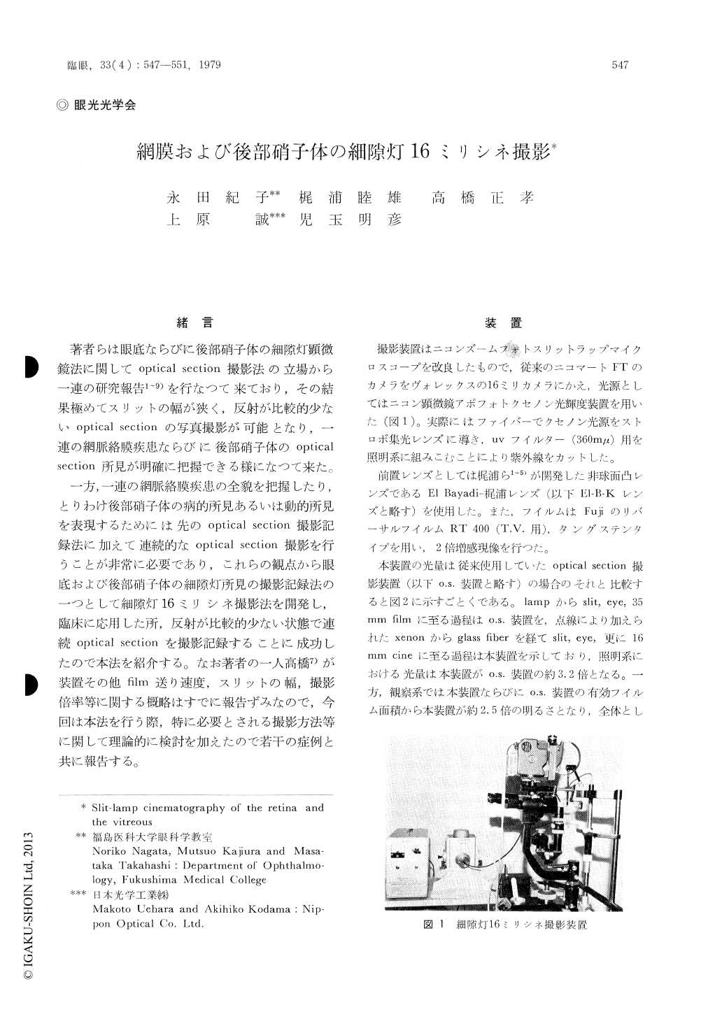
The authors recently developed a slit-lamp cinematography method for the retina and pos-terior vitreous body. The slit-lamp cinematogra-phy was devised by combining the Nikon apo-photo as light source and the Bolex 16mm camera mounted on the Nikon photo slit-lamp. The pre-set lens we used this time was the "El Bayadi-Kajiura lens". The illumination-observation angle required for the vitreous body is less than that required for the retina in photographing. In other words, the angle of admitting light into the retina is set under the same condition of the illumination-observation angle.

Copyright © 1979, Igaku-Shoin Ltd. All rights reserved.


