Japanese
English
- 有料閲覧
- Abstract 文献概要
- 1ページ目 Look Inside
緒 言
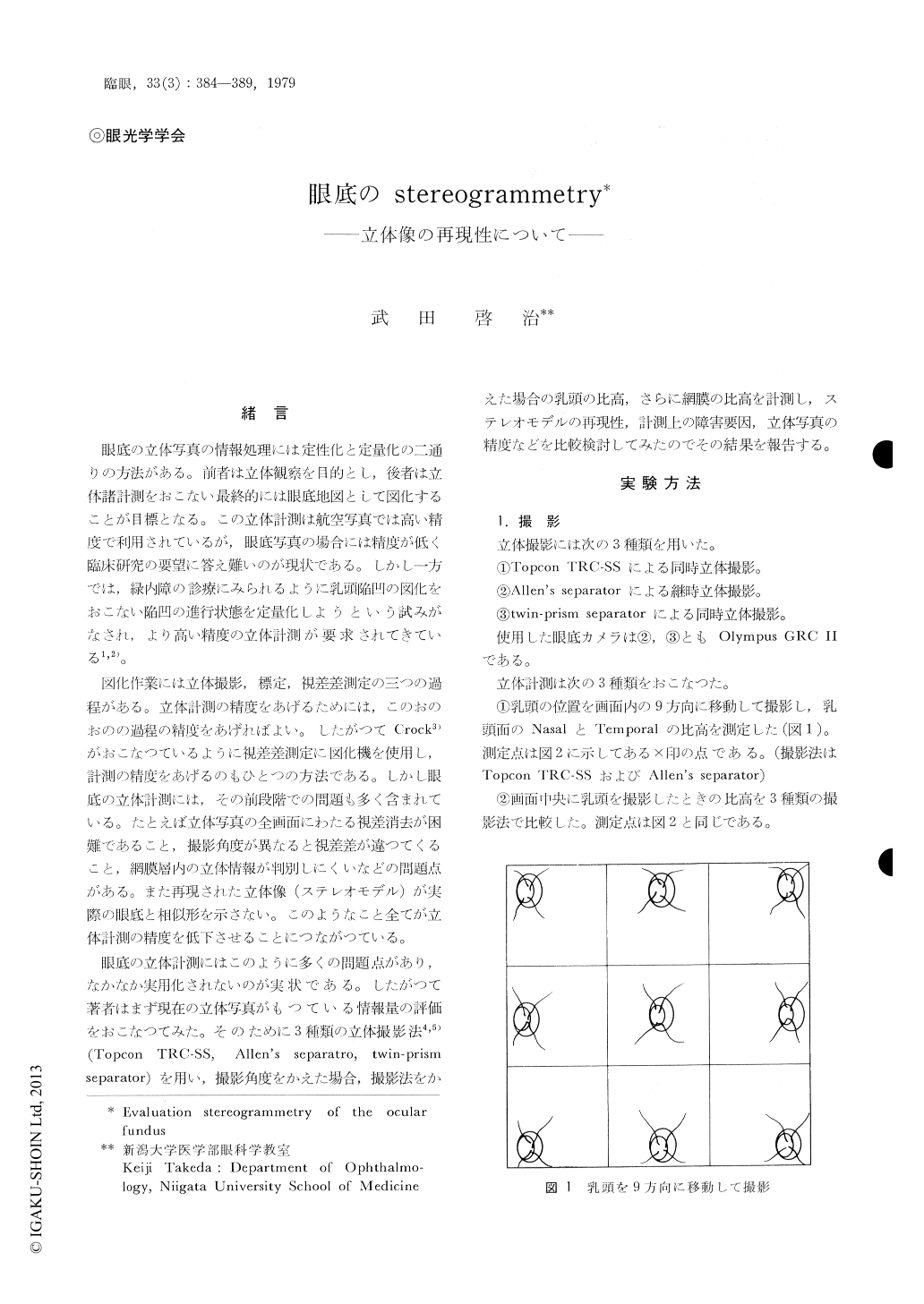
眼底の立体写真の情報処理には定性化と定量化の二通りの方法がある。前者は立体観察を目的とし,後者は立体諸計測をおこない最終的には眼底地図として図化することが目標となる。この立体計測は航空写真では高い精度で利用されているが,眼底写真の場合には精度が低く臨床研究の要望に答え難いのが現状である。しかし一方では,緑内障の診療にみられるように乳頭陥凹の図化をおこない陥凹の進行状態を定量化しようという試みがなされ,より高い精度の立体計測が要求されてきている1,2)。
図化作業には立体撮影,標定,視差差測定の三つの過程がある。立体計測の精度をあげるためには,このおのおのの過程の精度をあげればよい。したがつてCrock3)がおこなつているように視差差測定に図化機を使用し,計測の精度をあげるのもひとつの方法である。しかし眼底の立体計測には,その前段階での問題も多く含まれている。たとえば立体写真の全画面にわたる視差消去が困難であること,撮影角度が異なると視差差が違つてくること,網膜層内の立体情報が判別しにくいなどの問題点がある。また再現された立体像(ステレオモデル)が実際の眼底と相似形を示さない。このようなこと全てが立体計測の精度を低下させることにつながつている。
Stereo photogrammetry of the ocular fundus was studied by the use of the Topcon TRC-SS stereocamera, the Allen's separator method and the twin-prism separator method.
The parallux difference was measured by the mirror stereoscope and the stereometer (Nikon).
The height between the nasal and the tempo-ral margin of the optic disc and the thickness of the retina at the nasal margin of the disc were measured. Stereopairs of the optic disc were taken in the nine positions in the frame and were compaired one another to evaluate a positional error.
The result was as follows.

Copyright © 1979, Igaku-Shoin Ltd. All rights reserved.


