Japanese
English
- 有料閲覧
- Abstract 文献概要
- 1ページ目 Look Inside
緒 言
眼圧変動の影響による眼組織の変化にはいろいろなものがあり,そのうち高眼圧による変化は緑内障としてその病理学的検索はいうまでもなく螢光眼底撮影法などによる脈絡脈や視神経乳頭の循環動態の検討など現在もはなばなしく研究がなされているのは周知のところである。これに反し低眼圧の影響による眼組織の変化に関しては若干の文献が散見されるもののはなばなしい緑内障研究の陰にかくれあまり注目を集めていないのが実情である。しかし低眼圧によつてもたらされる種々の眼変化は日常臨床の場においても丹念に観察を行つているとかなりの頻度で遭遇する機会がある。すなわち眼外傷後の低眼圧,緑内障手術や白内障手術などの術後の低眼圧,網膜剥離眼にみられる低眼圧など身近かな症例においても低眼圧性眼変化が見出されうる。
低眼圧眼の臨床的所見について最初にかなりまとまつた報告をしているのはDellaporta1〜3),Pau4)であるが,その後永い間この問題は多くの研究者の関心を集めなかつた様である。近年に至りGass5)が螢光眼底撮影法を駆使し新しい解析を行い再び注目を集めだしたといえる。本邦においては横山6)がGassの説を紹介しその概要を述べているが,本邦ではHypotony maculopathyという言葉はまだ比較的馴染が薄い。
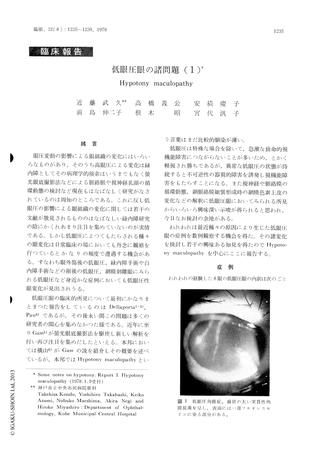
Observed 8 cases of hypotony maculopathy, in which hypotony was induced by glaucoma sur-gery (5 eyes), cyclodialysis (1), detachment of the ciliary epithelium (1) and penetrating corn-eal injury (1). The ocular hypotony was associat-ed with various clinical signs such as hypotony keratopathy, shallow anterior chamber, forward displacement of the lens, cataract formation, vitreous opacity, hypotony maculopathy and papilledema.
In hypotony maculopathy, the pattern of hypo-and hyperfluorescence in fluorescein angiograms did not always correspond to retinal folds as were observed ophthalmoscopically.

Copyright © 1978, Igaku-Shoin Ltd. All rights reserved.


