Japanese
English
- 有料閲覧
- Abstract 文献概要
- 1ページ目 Look Inside
緒 言
Ginestons1)の頃より,単色光による眼底所見の観察は多くの情報を提供するであろうとされていたが,1913年,Vogt2)により,初めて,その方法が実施された。その後,Francesch-ette & Müller(1930),Pavia(1931),Kleefeld(1935),Serr(1937),Ballentyne(1937)と報告が続いた。その結果は一定ではなく,臨床応用にには非実用的なものであつたが,眼底所見の把握に,新しい示唆を残した。単色光による眼底写真は,1934年,Kugelbergにより初めて発表されたが,当時,眼底カメラ,撮影手技,フイルムの感度,フイルターの特性等々,多くの問題があつた。
その後,フイルターの開発,眼底撮影法の向上に伴い,Potts3),Behrendt & Duane4),Beh-rendt & Wilson5),小島ら6)による詳細な分析は当時,注目を浴びた。PottsはTelevision ca-meraによる観察を行ない,Behrendtらは主波長が431,477,504,549,577,606および640nm,透過率40%,半値幅がほぼ10nmの干渉フイルターを用い,小島らはKodak Wrattenfilter No.17をおのおの使用し,眼底撮影を行なつた。
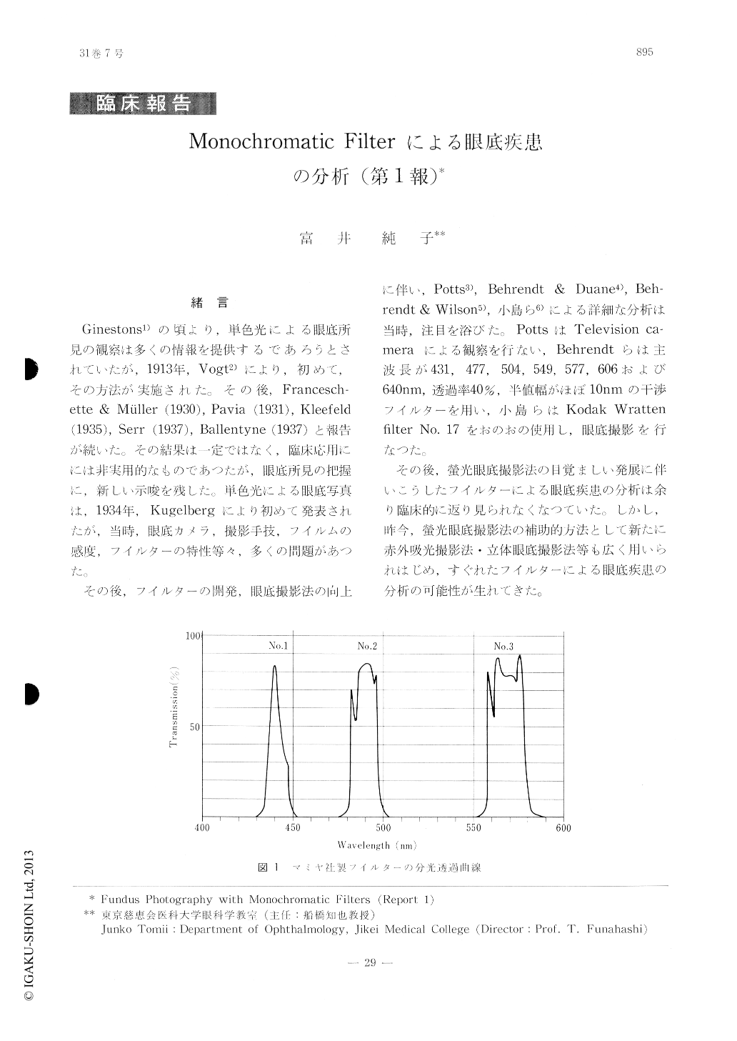
Normal and fundus fundi were photographed using three narrow-band monochromatic filters with transmission peaks at 440,490 and 570nm respectively. High-sensitive, B & W film was used for recording throughout.
The use of 570nm filter facilited recognition of paramacular pigment epithelial lesions and subretinal precipitates in central serous retino-pathy. It also facilitated recognition of neovas-cular membrane in deeper retinal layers in toxoplasmosis.

Copyright © 1977, Igaku-Shoin Ltd. All rights reserved.


