Japanese
English
- 有料閲覧
- Abstract 文献概要
- 1ページ目 Look Inside
緒 言
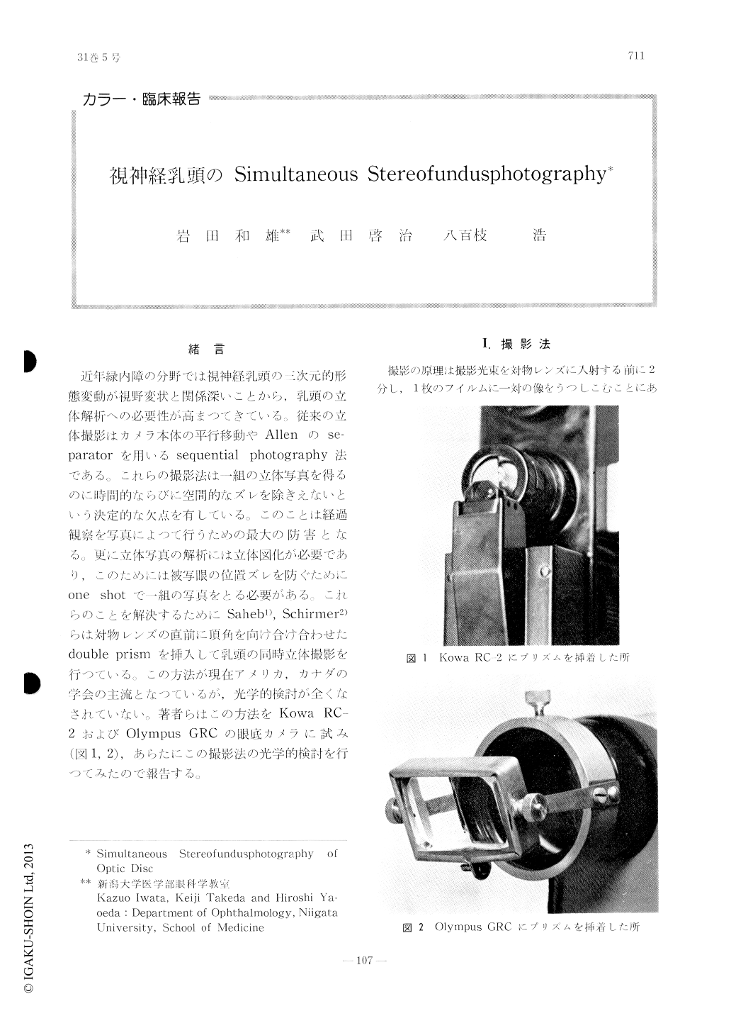
近年緑内障の分野では視神経乳頭の三次元的形態変動が視野変状と関係深いことから,乳頭の立体解析への必要性が高まつてきている.従来の立体撮影はカメラ本体の平行移動やAllenのse-Paratorを用いるsequential photograPhy法である。これらの撮影法は一組の立体写真を得るのに時間的ならびに空間的なズレを除きえないという決定的な欠点を有している。このことは経過観察を写真によつて行うための最大の防害となる。更に立体写真の解析には立体図化が必要であり,このためには被写眼の位置ズレを防ぐためにone shotで一組の写真をとる必要がある。これらのことを解決するためにSaheb1),Schinitier2)らは対物レンズの直前に頂角を向け合け合わせたdouble prismを挿入して乳頭の同時立体撮影を行つている。この方法が現在アメリカ,カナダの学会の主流となつているが,光学的検討が全くなされていない。著者らはこの方法をKowa RC-2およびOlympus GRCの眼底カメラに試み(図1,2),あらたにこの撮影法の光学的検討を行つてみたので報告する。
Simultaneous stereofundusphotograph of the optic disc was taken with a twin prism moun-ted in front of the objective of the fundus ca-mera. From our optical calculation a pair of 12△ prisms was optimum for Kowa RC-2 fun-dus camera and 10△ prisms for Olympus GRC fundus camera.
The distance of the separated two optic axis of the fundus camera at the entrance pupil of the eye, the distance of the twin images on the plane of the film were calculated, and the qu-ality of the twin pictures was optically analy-sed.

Copyright © 1977, Igaku-Shoin Ltd. All rights reserved.


